Explore
 Validate
 Learn
Learn Western blot
 Immunoprecipitation
Antibody data
- Antibody Data
- Antigen structure
- References [0]
- Comments [0]
- Validations
- Western blot [5]
- Immunocytochemistry [1]
- Immunohistochemistry [4]
Submit
Validation data
Reference
Comment
Report error
- Product number
- GTX22928 - Provider product page

- Provider
- GeneTex
- Proper citation
- GeneTex Cat#GTX22928, RRID:AB_384914
- Product name
- Hsp90 alpha antibody
- Antibody type
- Polyclonal
- Reactivity
- Human, Mouse, Rat, Rabbit, Sheep, Simian
- Host
- Rabbit
No comments: Submit comment
Supportive validation
- Submitted by
- GeneTex (provider)
- Main image

- Experimental details
- Western blot of mouse HSP 84using GTX22928.
- Submitted by
- GeneTex (provider)
- Main image

- Experimental details
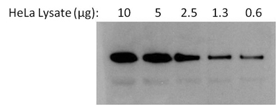
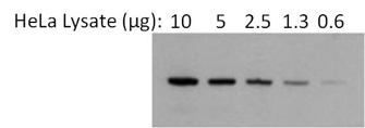
- Western blot analysis of Heat Shock Protein 86 (Hsp86) was performed by loading 2-fold serial dilutions of HeLa cell lysate, starting at 10 ug, per well onto a 4-20% Tris-HCl polyacrylamide gel. Proteins were transferred to a nitrocellulose membrane and blocked with 5% BSA/TBST for at least 1 hour. The membrane was probed with an Hsp86 polyclonal antibody (GTX22928) at a dilution of 1:1000 overnight at 4?C on a rocking platform, washed in TBS-0.1%Tween-20, and probed with an HRP-conjugated goat anti-rabbit IgG secondary antibody at a dilution of 1:10,000 for at least 1 hour. Chemiluminescent detection was performed using ECL Substrate.
- Submitted by
- GeneTex (provider)
- Main image
- Experimental details
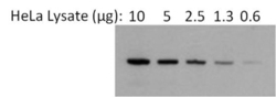
- Western blot analysis of Heat Shock Protein 86 (Hsp86) was performed by loading 2-fold serial dilutions of HeLa cell lysate, starting at 10 ug, per well onto a 4-20% Tris-HCl polyacrylamide gel. Proteins were transferred to a nitrocellulose membrane and blocked with 5% BSA/TBST for at least 1 hour. The membrane was probed with an Hsp86 polyclonal antibody (GTX22928) at a dilution of 1:1000 for 1 hour at room temperature on a rocking platform, washed in TBS-0.1%Tween-20, and probed with an AP-conjugated goat anti-rabbit IgG secondary antibody at a dilution of 1:10,000 for at least 1 hour. Chemiluminescent detection was performed using Lumi-Phos Substrate.
- Submitted by
- GeneTex (provider)
- Main image

- Experimental details
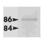
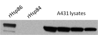
- Western blot analysis of epidermoid carcinoma (A431) cells using Heat Shock Protein 86 Polyclonal Antibody (GTX22928). Decreasing amounts of A431 whole cell lysates were probed with PA3-013 at a dilution of 1:2000 followed by an anti-rabbit IgG secondary antibody and SuperSignal West Pico Chemiluminescent Substrate. Lanes 1 and 2 are control samples of purified, recombinant Hsp86 (rHsp86) and Hsp84 (rHsp84), respectively. Data courtesy of the Innovators Program.
- Submitted by
- GeneTex (provider)
- Main image

- Experimental details
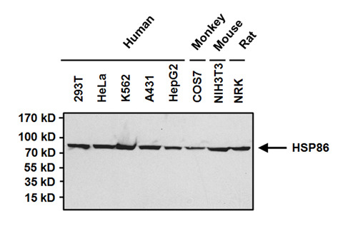
- Western blot analysis of Heat Shock Protein 86 (HSP86) was performed by loading 50ug of the indicated whole cell lysates and 15ul of PageRuler Prestained Protein Ladder onto a 4-20% Tris-HCl polyacrylamide gel. Proteins were transferred to a PVDF membrane and blocked with 5% BSA/TBST for at least 1 hour. The membrane was probed with a HSP86 polyclonal antibody (GTX22928) at a dilution of 1:1000 overnight at 4¢XC on a rocking platform, washed in TBS-0.1%Tween 20, and probed with a goat anti-rabbit IgG HRP secondary antibody at a dilution of 1:20,000 for at least 1 hour. Chemiluminescent detection was performed
Supportive validation
- Submitted by
- GeneTex (provider)
- Main image

- Experimental details
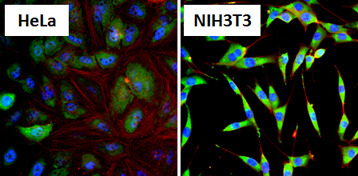
- Immunofluorescent analysis of Heat Shock Protein 86 (HSP86, green) in HeLa cells and NIH3T3 cells. Formalin fixed cells were permeabilized with 0.1% Triton X-100 in TBS for 10 minutes at room temperature and blocked with 1% Blocker BSA for 15 minutes at room temperature. Cells were probed with a HSP86 polyclonal antibody (GTX22928), at a dilution of 1:100 for at least 1 hour at room temperature, washed with PBS, and incubated with DyLight 488 goat-anti-rabbit IgG secondary antibody at a dilution of 1:400 for 30 minutes at room temperature. Nuclei (blue) were stained with Hoechst 33342 dye.
Supportive validation
- Submitted by
- GeneTex (provider)
- Main image

- Experimental details
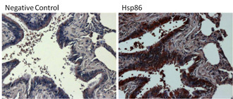
- Immunohistochemistry was performed on differentiated colon adenocarcinoma from paraffin-embedded human colon tissue sections. To expose target proteins, heat-induced epitope retrieval was performed using 10mM sodium citrate (pH 6.0) buffer for 20 minutes at 95?C. Following antigen retrieval, tissues were blocked in 3% BSA in PBST for 30 minutes at room temperature and then probed with a Heat Shock Protein 86 (Hsp86) polyclonal antibody (GTX22928) at a dilution of 1:100 for 1 hour in a humidified chamber (right panel). As a negative control, the primary antibody was eliminated from the staining procedure (left panel). Tissues were washed extensively with PBS/0.025% Tween-20 and endogenous peroxidase activity quenched with Peroxidase Suppressor for 30 minutes at room temperature. Detection was performed using an HRP-conjugated goat anti-rabbit IgG-HRP secondary antibody at a dilution of 1:250 followed by colorimetric detection using Metal Enhanced DAB Substrate Kit. Tissues were counterstained with hematoxylin and prepped for mounting.
- Submitted by
- GeneTex (provider)
- Main image
- Experimental details
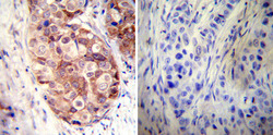
- Immunohistochemistry was performed on cancer biopsies of deparaffinized Human breast carcinoma tissues. To expose target proteins, heat induced antigen retrieval was performed using 10mM sodium citrate (pH6.0) buffer, microwaved for 8-15 minutes. Following antigen retrieval tissues were blocked in 3% BSA-PBS for 30 minutes at room temperature. Tissues were then probed at a dilution of 1:50 with a rabbit polyclonal antibody recognizing Heat Shock Protein 90 (86) (GTX22928) or without primary antibody (negative control) overnight at 4¢XC in a humidified chamber. Tissues were washed extensively with PBST and endogenous peroxidase activity was quenched with a peroxidase suppressor. Detection was performed using a biotin-conjugated secondary antibody and SA-HRP, followed by colorimetric detection using DAB. Tissues were counterstained with hematoxylin and prepped for mounting.

- Submitted by
- GeneTex (provider)
- Main image
- Experimental details
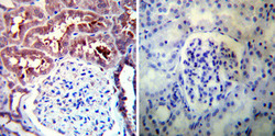
- Immunohistochemistry was performed on normal deparaffinized Human kidney tissue tissues. To expose target proteins, heat induced antigen retrieval was performed using 10mM sodium citrate (pH6.0) buffer, microwaved for 8-15 minutes. Following antigen retrieval tissues were blocked in 3% BSA-PBS for 30 minutes at room temperature. Tissues were then probed at a dilution of 1:100 with a rabbit polyclonal antibody recognizing Heat Shock Protein 90 (86) (GTX22928) or without primary antibody (negative control) overnight at 4¢XC in a humidified chamber. Tissues were washed extensively with PBST and endogenous peroxidase activity was quenched with a peroxidase suppressor. Detection was performed using a biotin-conjugated secondary antibody and SA-HRP, followed by colorimetric detection using DAB. Tissues were counterstained with hematoxylin and prepped for mounting.

- Submitted by
- GeneTex (provider)
- Main image

- Experimental details
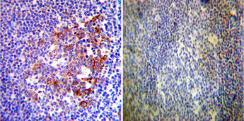
- Immunohistochemistry was performed on normal deparaffinized Human tonsil tissue tissues. To expose target proteins, heat induced antigen retrieval was performed using 10mM sodium citrate (pH6.0) buffer, microwaved for 8-15 minutes. Following antigen retrieval tissues were blocked in 3% BSA-PBS for 30 minutes at room temperature. Tissues were then probed at a dilution of 1:200 with a rabbit polyclonal antibody recognizing Heat Shock Protein 90 (86) (GTX22928) or without primary antibody (negative control) overnight at 4¢XC in a humidified chamber. Tissues were washed extensively with PBST and endogenous peroxidase activity was quenched with a peroxidase suppressor. Detection was performed using a biotin-conjugated secondary antibody and SA-HRP, followed by colorimetric detection using DAB. Tissues were counterstained with hematoxylin and prepped for mounting.