Explore
 Validate
 Learn
Learn Western blot
Antibody data
- Antibody Data
- Antigen structure
- References [0]
- Comments [0]
- Validations
- Western blot [4]
- Immunohistochemistry [1]
Submit
Validation data
Reference
Comment
Report error
- Product number
- PA5-46936 - Provider product page

- Provider
- Invitrogen Antibodies
- Product name
- Calbindin D28K Polyclonal Antibody
- Antibody type
- Polyclonal
- Antigen
- Recombinant full-length protein
- Description
- Reconstitute at 0.2 mg/mL in sterile PBS.
- Reactivity
- Human
- Host
- Goat
- Isotype
- IgG
- Vial size
- 100 µg
- Concentration
- 0.2 mg/mL
- Storage
- -20° C, Avoid Freeze/Thaw Cycles
No comments: Submit comment
Supportive validation
- Submitted by
- Invitrogen Antibodies (provider)
- Main image
- Experimental details
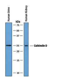
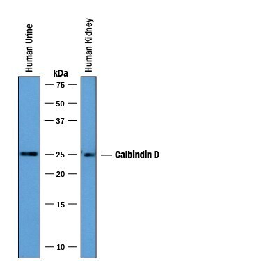
- Western blot analysis from human urine and lysates of human kidney tissue. PVDF membrane was probed with 5 µg/mL of Goat Anti-human Calbindin D Antigen Affinity-purified Polyclonal Antibody (Product # PA5-46936) followed by HRP-conjugated Anti-Goat IgG Secondary Antibody. A specific band was detected for Calbindin D at approximately 25 kDa (as indicated). This experiment was conducted under reducing conditions.

- Submitted by
- Invitrogen Antibodies (provider)
- Main image
- Experimental details
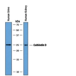
- Western blot analysis of Calbindin D28K in human urine and lysates of human kidney tissue. Samples were incubated in Calbindin D28K polyclonal antibody (Product # PA5-46936) using a dilution of 5 µg/mL followed by a HRP-conjugated Anti-Goat IgG secondary antibody. A specific band was detected for Calbindin D at approximately 25 kDa (as indicated). This experiment was conducted under reducing conditions.
- Submitted by
- Invitrogen Antibodies (provider)
- Main image
- Experimental details
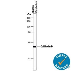
- Western blot analysis of Calbindin D28K in 0.2 mg/mL lysates of human brain (cerebellum) tissue. Samples were incubated in Calbindin D28K polyclonal antibody (Product # PA5-46936) at a dilution of 10 µg/mL. A specific band was detected for Calbindin D at approximately 32 kDa (as indicated). This experiment was conducted under reducing conditions and using the 12-230 kDa separation system.

- Submitted by
- Invitrogen Antibodies (provider)
- Main image

- Experimental details
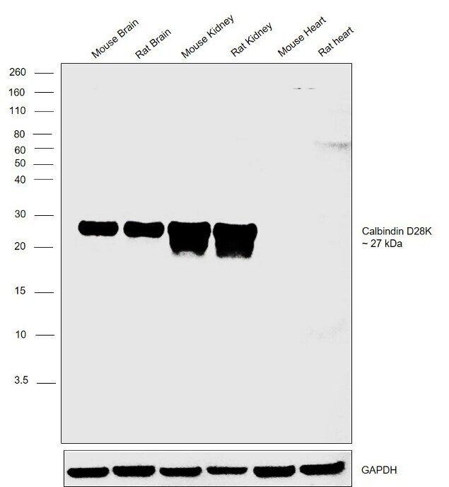
- Western blot was performed using Anti-Calbindin D28K Polyclonal Antibody (Product # PA5-46936) and a 27 kDa band corresponding to Calbindin D28K was observed across tissues tested except Mouse and Rat Heart which are reported to be negative. Whole cell extracts (30 µg lysate) of Mouse Brain (Lane 1), Rat Brain (Lane 2), Mouse Kidney (Lane 3), Rat Kidney (Lane 4), Mouse Heart (Lane 5) and Rat Heart (Lane 6) were electrophoresed using Novex® NuPAGE® 4-12 % Bis-Tris gel (Product # NP0321BOX). Resolved proteins were then transferred onto a nitrocellulose membrane (Product # IB23001) by iBlot® 2 Dry Blotting System (Product # IB21001). The blot was probed with the primary antibody (5 ug/ml) and detected by chemiluminescence with Rabbit anti-Goat IgG (H+L), Superclonal™ Recombinant Secondary Antibody, HRP (Product # A27014, 1:4000 dilution) using the iBright FL 1000 (Product # A32752). Chemiluminescent detection was performed using Novex® ECL Chemiluminescent Substrate Reagent Kit (Product # WP20005).
Supportive validation
- Submitted by
- Invitrogen Antibodies (provider)
- Main image
- Experimental details
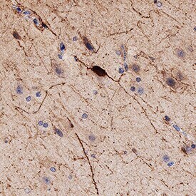
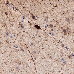
- Immunohistochemical analysis of Calbindin D28K in immersion fixed paraffin-embedded sections of human brain (hippocampus). Samples were incubated with Calbindin D28K polyclonal antibody (Product # PA5-46936) using a dilution of 3 µg/mL for 1 hour at room temperature followed by Anti-Goat IgG VisUCyte™ HRP Polymer Antibody. Before incubation with the primary antibody, tissue was subjected to heat-induced epitope retrieval using Antigen Retrieval Reagent-Basic . Tissue was stained using DAB (brown) and counterstained with hematoxylin (blue). Specific staining was localized to neuronal cell bodies and synaptic vesicles.