Explore
 Validate
 Learn
Learn Western blot
 Flow cytometry
Antibody data
- Antibody Data
- Antigen structure
- References [0]
- Comments [0]
- Validations
- Flow cytometry [4]
Submit
Validation data
Reference
Comment
Report error
- Product number
- NBP1-48348DL594 - Provider product page

- Provider
- Novus Biologicals
- Product name
- Mouse Monoclonal Cytokeratin, pan Antibody
- Antibody type
- Monoclonal
- Description
- Protein A purified. This Mouse monoclonal Cytokeratin (Pan-reactive) antibody reacts with Cytokeratin peptides 4, 5, 6, 8, 10, 13, 18. Cytokeratins are a member of intermediate filaments subfamily represented in epithelial tissues.
- Reactivity
- Human, Mouse, Rat
- Host
- Mouse
- Conjugate
- Red dye
- Isotype
- IgG
- Vial size
- 0.1 ml
- Concentration
- 1 mg/ml
- Storage
- Store at 4C short term. For extended storage, add an equal volume of glycerol, aliquot and store at -20C or below. Avoid repeated freeze-thaw cycles.
No comments: Submit comment
Supportive validation
- Submitted by
- Novus Biologicals (provider)
- Main image
- Experimental details
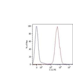
- Flow Cytometry: pan Cytokeratin Antibody (C-11) [DyLight 594] [NBP1-48348DL594] - Intracellular flow cytometry analysis of cytokeratin expression in HT-29 human Caucasian colon adenocarcinoma cell line using anti-cytokeratin antibody (C-11) PE. Overlay with Isotype mouse IgG1 control.
- Submitted by
- Novus Biologicals (provider)
- Main image

- Experimental details
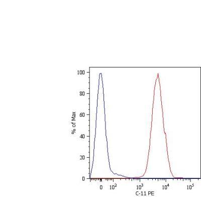
- Flow Cytometry: pan Cytokeratin Antibody (C-11) [DyLight 594] [NBP1-48348DL594] - Intracellular flow cytometry analysis of cytokeratin expression in HeLa cells.
- Submitted by
- Novus Biologicals (provider)
- Main image
- Experimental details
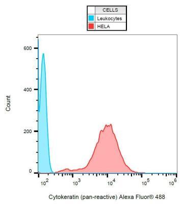
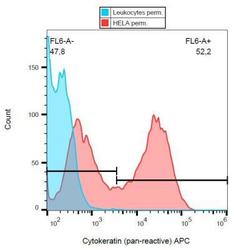
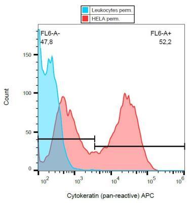
- Flow Cytometry: pan Cytokeratin Antibody (C-11) [DyLight 594] [NBP1-48348DL594] - Intracellular flow cytometry analysis of cytokeratin expression in HeLa cells using the APC conjugate.
- Submitted by
- Novus Biologicals (provider)
- Main image

- Experimental details
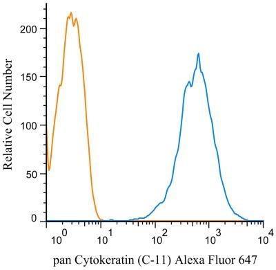
- Flow (Intracellular): pan Cytokeratin Antibody (C-11) [DyLight 594] [NBP1-48348DL594] - An intracellular stain was performed on HeLa cells with pan Cytokeratin antibody (C-11) NBP1-48348AF647 (blue) and a matched isotype control NBP2-27287AF647 (orange). Cells were fixed with 4% PFA and then permeablized with 0.1% saponin. Cells were incubated in an antibody dilution of 2 ug/mL for 30 minutes at room temperature. Both antibodies were conjugated to Alexa Fluor 647.