 Explore
 Validate
 Learn
Learn Immunohistochemistry
Antibody data
- Antibody Data
- Antigen structure
- References [3]
- Comments [0]
- Validations
- Immunohistochemistry [1]
- Other assay [1]
Submit
Validation data
Reference
Comment
Report error
- Product number
- PA1-27114 - Provider product page

- Provider
- Invitrogen Antibodies
- Product name
- Cytokeratin Pan Polyclonal Antibody
- Antibody type
- Polyclonal
- Antigen
- Other
- Description
- This antibody will detect Cytokeratins 4, 5, 6, (and to a lesser extent) 8, 14, and 16.
- Reactivity
- Human, Bovine
- Host
- Rabbit
- Isotype
- IgG
- Vial size
- 500 µL
- Storage
- 4° C, do not freeze
Submitted references Exposure to Microbial Metabolite Butyrate Prolongs the Survival Time and Changes the Growth Pattern of Human Papillomavirus 16 E6/E7-Immortalized Keratinocytes in Vivo.
Bioprinted Skin Recapitulates Normal Collagen Remodeling in Full-Thickness Wounds.
Separation of breast cancer and organ microenvironment transcriptomes in metastases.
Li M, McGhee EM, Shinno L, Lee K, Lin YL
The American journal of pathology 2021 Oct;191(10):1822-1836
The American journal of pathology 2021 Oct;191(10):1822-1836
Bioprinted Skin Recapitulates Normal Collagen Remodeling in Full-Thickness Wounds.
Jorgensen AM, Varkey M, Gorkun A, Clouse C, Xu L, Chou Z, Murphy SV, Molnar J, Lee SJ, Yoo JJ, Soker S, Atala A
Tissue engineering. Part A 2020 May;26(9-10):512-526
Tissue engineering. Part A 2020 May;26(9-10):512-526
Separation of breast cancer and organ microenvironment transcriptomes in metastases.
Alzubi MA, Turner TH, Olex AL, Sohal SS, Tobin NP, Recio SG, Bergh J, Hatschek T, Parker JS, Sartorius CA, Perou CM, Dozmorov MG, Harrell JC
Breast cancer research : BCR 2019 Mar 6;21(1):36
Breast cancer research : BCR 2019 Mar 6;21(1):36
No comments: Submit comment
Supportive validation
- Submitted by
- Invitrogen Antibodies (provider)
- Main image

- Experimental details
- Immunohistochemical (Paraffin) analysis of human skin tissue using (Product # PA1-27114) Cytokeratin Pan Polyclonal Antibody.

Supportive validation
- Submitted by
- Invitrogen Antibodies (provider)
- Main image
- Experimental details
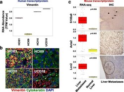
- Fig. 4 Validation of human and mouse-specific transcripts with immunofluorescence microscopy and immunohistochemistry. a Box and whisker plots showing human vimentin RNA expression levels in TNBC mammary tumors. b Immunofluorescence microscopy for pan-cytokeratin (green), vimentin (red), and DAPI (blue) in TNBC mammary tumors. c The mouse RNA-seq dataset was queried for genes that were differentially expressed in normal mouse liver as compared to mouse livers colonized by metastatic cells. Immunohistochemistry of liver metastases to validate the increased RNA expression observed in liver metastases. Asterisk denotes the location of cancer cells; arrows denote S100a9 cells in the peri-tumor area surrounding the cancer cells
