 Explore
 Validate
 Learn
Learn Flow cytometry
Antibody data
- Antibody Data
- Antigen structure
- References [5]
- Comments [0]
- Validations
- Flow cytometry [1]
Submit
Validation data
Reference
Comment
Report error
- Product number
- 25-0839-41 - Provider product page

- Provider
- Invitrogen Antibodies
- Product name
- Anti-CD83 Monoclonal Antibody (HB15e), PE-Cyanine7, eBioscience™
- Antibody type
- Monoclonal
- Antigen
- Other
- Description
- Description: The HB15e monoclonal antibody reacts with human CD83, a 45 kDa transmembrane glycoprotein. CD83, a member of the Ig superfamily, is expressed on cultured dendritic cells, interdigitating, follicular, and circulating dendritic cells as well as some proliferating lymphocytes, and human cell lines express this antigen. While the function of CD83 is unclear, it can serve as a useful marker for mature human blood dendritic cells. Applications Reported: This HB15e antibody has been reported for use in flow cytometric analysis. Applications Tested: This HB15e antibody has been pre-titrated and tested by flow cytometric analysis of normal human peripheral blood cells. This can be used at 5 µL (0.25 µg) per test. A test is defined as the amount (µg) of antibody that will stain a cell sample in a final volume of 100 µL. Cell number should be determined empirically but can range from 10^5 to 10^8 cells/test. Light sensitivity: This tandem dye is sensitive photo-induced oxidation. Please protect this vial and stained samples from light. Fixation: Samples can be stored in IC Fixation Buffer (cat. 00-8222) (100 µL cell sample + 100 µL IC Fixation Buffer) or 1-step Fix/Lyse Solution (cat. 00-5333) for up to 3 days in the dark at 4°C with minimal impact on brightness and FRET efficiency/compensation. Some generalizations regarding fluorophore performance after fixation can be made, but clone specific performance should be determined empirically. Excitation: 488-561 nm; Emission: 775 nm; Laser: Blue Laser, Green Laser, Yellow-Green Laser. Filtration: 0.2 µm post-manufacturing filtered.
- Reactivity
- Human
- Host
- Mouse
- Isotype
- IgG
- Antibody clone number
- HB15e
- Vial size
- 25 Tests
- Concentration
- 5 µL/Test
- Storage
- 4° C, store in dark, DO NOT FREEZE!
Submitted references Schistosoma mansoni rSm29 Antigen Induces a Regulatory Phenotype on Dendritic Cells and Lymphocytes From Patients With Cutaneous Leishmaniasis.
Surface LAMP-2 Is an Endocytic Receptor That Diverts Antigen Internalized by Human Dendritic Cells into Highly Immunogenic Exosomes.
Dendritic cell profile induced by Schistosoma mansoni antigen in cutaneous leishmaniasis patients.
Reprogramming human endothelial cells to haematopoietic cells requires vascular induction.
Noncanonical dendritic cell differentiation and survival driven by a bacteremic pathogen.
Lopes DM, Oliveira SC, Page B, Carvalho LP, Carvalho EM, Cardoso LS
Frontiers in immunology 2018;9:3122
Frontiers in immunology 2018;9:3122
Surface LAMP-2 Is an Endocytic Receptor That Diverts Antigen Internalized by Human Dendritic Cells into Highly Immunogenic Exosomes.
Leone DA, Peschel A, Brown M, Schachner H, Ball MJ, Gyuraszova M, Salzer-Muhar U, Fukuda M, Vizzardelli C, Bohle B, Rees AJ, Kain R
Journal of immunology (Baltimore, Md. : 1950) 2017 Jul 15;199(2):531-546
Journal of immunology (Baltimore, Md. : 1950) 2017 Jul 15;199(2):531-546
Dendritic cell profile induced by Schistosoma mansoni antigen in cutaneous leishmaniasis patients.
Lopes DM, Fernandes JS, Cardoso TM, Bafica AM, Oliveira SC, Carvalho EM, Araujo MI, Cardoso LS
BioMed research international 2014;2014:743069
BioMed research international 2014;2014:743069
Reprogramming human endothelial cells to haematopoietic cells requires vascular induction.
Sandler VM, Lis R, Liu Y, Kedem A, James D, Elemento O, Butler JM, Scandura JM, Rafii S
Nature 2014 Jul 17;511(7509):312-8
Nature 2014 Jul 17;511(7509):312-8
Noncanonical dendritic cell differentiation and survival driven by a bacteremic pathogen.
Miles B, Scisci E, Carrion J, Sabino GJ, Genco CA, Cutler CW
Journal of leukocyte biology 2013 Aug;94(2):281-9
Journal of leukocyte biology 2013 Aug;94(2):281-9
No comments: Submit comment
Supportive validation
- Submitted by
- Invitrogen Antibodies (provider)
- Main image
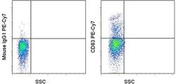
- Experimental details
- Staining of 1-day PHA stimulated normal human peripheral blood cells with Mouse IgG1 K Isotype Control PE-Cyanine7 (Product # 25-4714-80) (left) or Anti-Human CD83 PE-Cyanine7 (right). Total viable cells were used for analysis.
