Explore
 Validate
 Learn
Learn Western blot
Antibody data
- Antibody Data
- Antigen structure
- References [0]
- Comments [0]
- Validations
- Western blot [2]
- Immunocytochemistry [2]
- Immunohistochemistry [1]
- Flow cytometry [1]
Submit
Validation data
Reference
Comment
Report error
- Product number
- 701949 - Provider product page

- Provider
- Invitrogen Antibodies
- Product name
- Tyrosine Hydroxylase Recombinant Rabbit Monoclonal Antibody (9H8L15)
- Antibody type
- Monoclonal
- Antigen
- Other
- Description
- This antibody is predicted to react with Monkey and Pig.
- Antibody clone number
- 9H8L15
- Concentration
- 0.5 mg/mL
No comments: Submit comment
Supportive validation
- Submitted by
- Invitrogen Antibodies (provider)
- Main image
- Experimental details
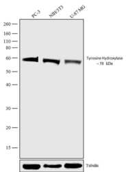
- Western blot analysis was performed on whole cell extracts (30 µg lysate) of PC-3 (Lane 1), NIH/3T3 (Lane 2), U-87MG (Lane 3). The blots were probed with Anti-Tyrosine Hydroxylase Recombinant Rabbit Monoclonal Antibody (Product # 701949, 1-2 µg/mL) and detected by chemiluminescence using Goat anti-Rabbit IgG (H+L) Superclonal™ Secondary Antibody, HRP conjugate (Product # A27036, 0.4 µg/mL, 1:2500 dilution). A 58 kDa band corresponding to Tyrosine Hydroxylase was observed. Known quantity of protein samples were electrophoresed using Novex® NuPAGE® 10% Bis-Tris gel (Product # NP0301BOX), XCell SureLock™ Electrophoresis System (Product # EI0002) and Novex® Sharp Pre-Stained Protein Standard (Product # LC5800). Resolved proteins were then transferred onto a nitrocellulose membrane with iBlot® Dry Blotting System (Product # IB21001). The membrane was probed with the relevant primary and secondary Antibody following blocking with 5% skimmed milk. Chemiluminescent detection was performed using Pierce™ ECL Western blotting Substrate (Product # 32106).

- Submitted by
- Invitrogen Antibodies (provider)
- Main image
- Experimental details
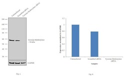
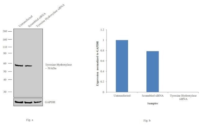
- Knockdown of Tyrosine Hydroxylase was achieved by transfecting Caco-2 with Tyrosine Hydroxylase specific siRNAs (Silencer® select Product # s14094). Western blot analysis (Fig. a) was performed using whole cell extracts from the Tyrosine Hydroxylase knockdown cells (lane 3), non-specific scrambled siRNA transfected cells (lane 2) and untransfected cells (lane 1). The blots were probed with Tyrosine Hydroxylase Antibody (9H8L15) (Product # 701949, 1 µg/ml) and Goat anti-Rabbit IgG (H+L) Superclonal™ Secondary Antibody, HRP conjugate (Product # A27036, 0.25µg/ml, 1:4000 dilution). Densitometric analysis of this western blot is shown in histogram (Fig. b). Decrease in signal upon siRNA mediated knock down confirms that antibody is specific to Tyrosine Hydroxylase.
Supportive validation
- Submitted by
- Invitrogen Antibodies (provider)
- Main image
- Experimental details
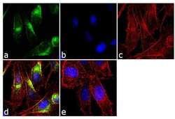
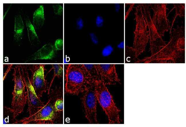
- Immunofluorescence was performed on fixed and permeabilized PC-3 cells for detection of TH using Anti-TH Recombinant Rabbit Monoclonal Antibody (Product # 701949, 2 µg/mL) and labeled with Goat anti-Rabbit IgG (H+L) Superclonal™ Secondary Antibody, Alexa Fluor® 488 conjugate (Product # A27034, 1:2000). Panel a) shows representative cells that were stained for detection and localization of TH protein (green), Panel b) is stained for nuclei (blue) using SlowFade® Gold Antifade Mountant with DAPI (Product # S36938). Panel c) represents cytoskeletal F-actin staining using Alexa Fluor® 555 Rhodamine Phalloidin (Product # R415, 1:300). Panel d) is a composite image of Panels a, b and c clearly demonstrating cytoplasmic localization of RNF20. Panel e) represents control cells with no primary antibody to assess background.
- Submitted by
- Invitrogen Antibodies (provider)
- Main image
- Experimental details
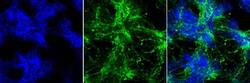
- Immunofluorescence analysis of DA neurons using anti-tyrosine hydroxylase (TH) antibody. H9 ESCs were differentiated with PSC Dopaminergic neuron differentiation kit (Product # A3147701). Expression of TH was labeled with TH monoclonal antibody (Product # 701949) followed by 2nd antibody AlexaFluor488 donkey anti-rabbit (Product # A-21206, green). Nuclear DNA was stained with DAPI (blue).

Supportive validation
- Submitted by
- Invitrogen Antibodies (provider)
- Main image

- Experimental details
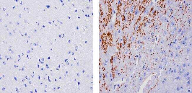
- Immunohistochemistry analysis of Tyrosine Hydroxylase showing staining in the Cytoplasm/Nerve fiber of paraffin-embedded mouse brain tissue (right) compared to a negative control without primary antibody (left). To expose target proteins, antigen retrieval was performed using 10mM sodium citrate (pH 6.0), microwaved for 8-15 min. Following antigen retrieval, tissues were blocked in 3% H2O2-methanol for 15 min at room temperature, washed with ddH2O and PBS, and then probed with a Tyrosine Hydroxylase Recombinant Rabbit Monoclonal Antibody (Product # 701949) diluted in 3% BSA-PBS at a dilution of 1:100 for 1 hour at 37ºC in a humidified chamber. Tissues were washed extensively in PBST and detection was performed using an HRP-conjugated secondary antibody followed by colorimetric detection using a DAB kit. Tissues were counterstained with hematoxylin and dehydrated with ethanol and xylene to prep for mounting.
Supportive validation
- Submitted by
- Invitrogen Antibodies (provider)
- Main image
- Experimental details
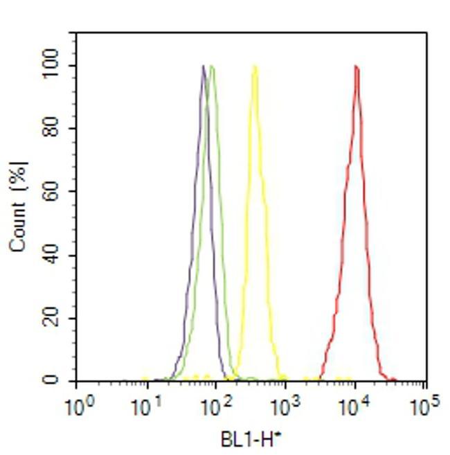
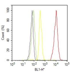
- Flow Cytometry analysis of TH was performed on PC-12 cells labeled with ABfinity™ Anti-TH Recombinant Rabbit Monoclonal Antibody (Product# 701949, 2-4 ug/ 1M cells) or with Rabbit isotype control and detected with Goat anti-Rabbit IgG (H+L) Superclonal™ Secondary Antibody, (Alexa Fluor® 488 conjugate, Product # A27034, 0.4 ug/ml, 1:2500) as represented by the red and yellow histograms respectively. The purple histogram represents unstained control cells and the green histogram represents no-primary-Antibody control. A representative of 10,000 cells were acquired and analyzed for each sample using an Attune® Acoustic Focusing Cytometer (4468770).