Explore
 Validate
 Learn
Learn Western blot
Antibody data
- Antibody Data
- Antigen structure
- References [0]
- Comments [0]
- Validations
- Western blot [2]
- Immunocytochemistry [1]
- Immunohistochemistry [1]
Submit
Validation data
Reference
Comment
Report error
- Product number
- PA5-18372 - Provider product page

- Provider
- Invitrogen Antibodies
- Product name
- Tyrosine Hydroxylase Polyclonal Antibody
- Antibody type
- Polyclonal
- Antigen
- Synthetic peptide
- Description
- This antibody is predicted to react with canine and rat based on sequence homology.
- Reactivity
- Human
- Host
- Goat
- Isotype
- IgG
- Vial size
- 100 µg
- Concentration
- 0.5 mg/mL
- Storage
- -20° C, Avoid Freeze/Thaw Cycles
No comments: Submit comment
Supportive validation
- Submitted by
- Invitrogen Antibodies (provider)
- Main image

- Experimental details
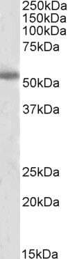
- Western Blot staining of Human Cerebral Cortex lysate using Product # PA5-18372 at a concentration of 2 µg/mL, the primary antibody incubation was 1 hour and the detection method was chemiluminescence.
- Submitted by
- Invitrogen Antibodies (provider)
- Main image

- Experimental details
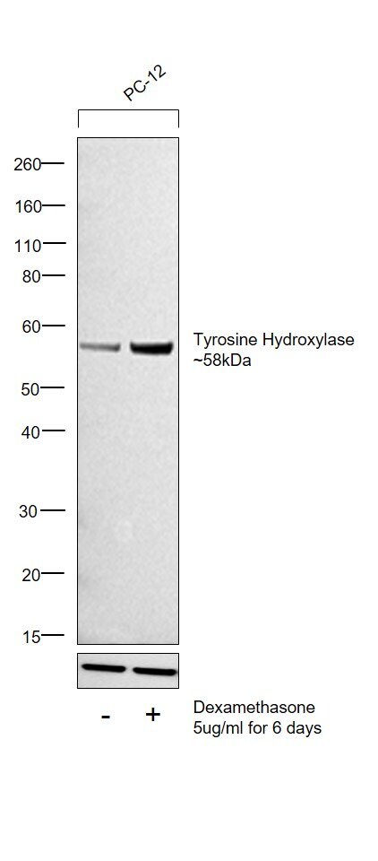
- Western blot was performed using Anti-Tyrosine Hydroxylase Polyclonal Antibody (Product # PA5-18372) and a 58kDa band corresponding to Tyrosine Hydroxylase was observed in PC-12 and was upregulated upon treatment with Dexamethasone. Whole Cell Extract-WCL (10 µg lysate) of PC-12 (Lane 1) and PC-12 treated with Dexamethasone (5ug/ml for 6 days) (Lane 2) were electrophoresed using NuPAGE™ 4-12% Bis-Tris Protein Gel (Product # NP0321BOX). Resolved proteins were then transferred onto a Nitrocellulose membrane (Product # IB23001) by iBlot® 2 Dry Blotting System (Product # IB21001). The blot was probed with the primary antibody (0.5ug/ml dilution) and detected by chemiluminescence with Rabbit anti-Goat IgG (H+L) Superclonal™ Recombinant Secondary Antibody, HRP (Product # A27014, 1:4000 dilution) using the iBright FL 1000 (Product # A32752). Chemiluminescent detection was performed using Novex® ECL Chemiluminescent Substrate Reagent Kit (Product # WP20005).
Supportive validation
- Submitted by
- Invitrogen Antibodies (provider)
- Main image
- Experimental details
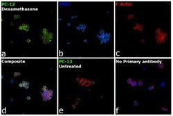
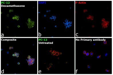
- Immunofluorescence analysis of Tyrosine Hydroxylase was performed using 70% confluent log phase PC-12 cells treated with Dexamethasone (5 µg/mL, 6 days). The cells were fixed with 4% paraformaldehyde for 10 minutes, permeabilized with 0.1% Triton™ X-100 for 15 minutes, and blocked with 2% BSA for 45 minutes at room temperature. The cells were labeled with Tyrosine Hydroxylase Polyclonal Antibody (Product # PA5-18372) at 1:100 dilution in 0.1% BSA, incubated at 4 degree celsius overnight and then labeled with Donkey anti-Goat IgG (H+L) Highly Cross-Adsorbed Secondary Antibody, Alexa Fluor Plus 488 (Product # A32814), (1:2000 dilution), for 45 minutes at room temperature (Panel a: Green). Nuclei (Panel b:Blue) were stained with ProLong™ Diamond Antifade Mountant with DAPI (Product # P36962). F-actin (Panel c: Red) was stained with Rhodamine Phalloidin (Product # R415, 1:300 dilution). Panel d represents the merged image showing increased expression of Tyrosine Hydroxylase upon treatment with Dexamethasone. Panel e represents untreated PC-12 cells with low expression of Tyrosine Hydroxylase. Panel f represents control cells with no primary antibody to assess background. The images were captured at 60x magnification.
Supportive validation
- Submitted by
- Invitrogen Antibodies (provider)
- Main image
- Experimental details
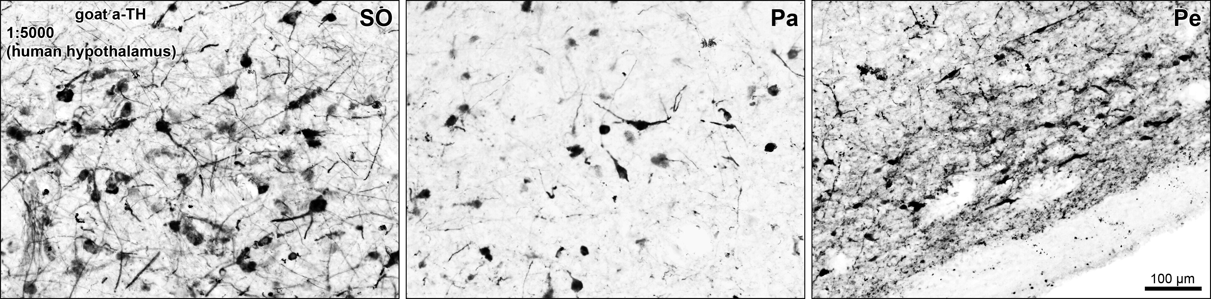
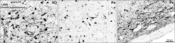
- Immunohistochemical analysis of Tyrosine Hydroxylase in Human hypothalamus using a Tyrosine Hydroxylase monoclonal antibody (Product #PA5-18677). The Human Hypothalamic supraoptic (SO), paraventricular (Pa) and periventricular (Pe) nuclei tissue sections were detected using antigen retrieval with citrate buffer pH 6, at 80C for 30min, followed by HRP-staining with Ni-DAB after Biotin-SP-antigoat amplification.