Explore
 Validate
 Learn
Learn Western blot
Antibody data
- Antibody Data
- Antigen structure
- References [0]
- Comments [0]
- Validations
- Western blot [1]
- Immunocytochemistry [2]
Submit
Validation data
Reference
Comment
Report error
- Product number
- ABIN2509236 - Provider product page

- Provider
- antibodies-online
- Product name
- anti-Chemokine (C-C Motif) Ligand 17 (CCL17) antibody
- Antibody type
- Polyclonal
- Antigen
- Other
- Description
- Produced from sera of rabbits pre-immunized with highly pure (>98%) recombinant hTARC. Anti-Human TARC specific antibody was purified by affinity chromatography employing immobilized hTARC matrix.
- Reactivity
- Human
- Host
- Rabbit
- Vial size
- 100 μg
- Storage
- -20°C
No comments: Submit comment
Supportive validation
- Submitted by
- antibodies-online (provider)
- Main image

- Experimental details
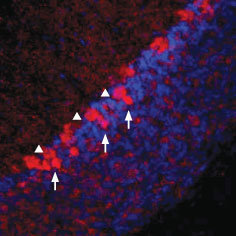
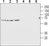
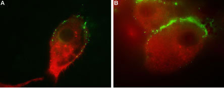
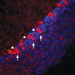
- Western blot analysis of rat brain membranes (lanes 1 and 4), mouse brain membranes (lanes 2 and 5) and human U-87 MG glyoblastoma lysates (lanes 3 and 6): 1-3. Anti-Vesicular GABA Transporter antibody (ABIN2511175), (1:200). 4-6. Anti-Vesicular GABA Transporter antibody, preincubated with the control peptide antigen. Expression of Vesicular GABA Transporter in rat cerebellum Immunohistochemical staining of rat frozen cerebellum sections using Anti-Vesicular GABA Transporter antibody (ABIN2511175), (1:100). VGAT staining is shown in red and dapi (blue) was used as a general cellular marker. VGAT appears in the cerebellar pinceau (arrows) and in the soma of Purkinje cells (triangles). Colocalization of AMPA Receptor 1 (GluR1) and Vesicular GABA Transporter in human U-87 MG cells Immunocytochemical staining of human glioblastoma U-87 MG. Extracellular staining of live intact cells with Anti-AMPA Receptor 1 (GluR1) (extracellular) antibody (AGP-009), (1:25), followed by goat anti-guinea pig-AlexaFluor-488 secondary antibody (green). Cells were subsequently fixed, permeabilized and labeled with Anti-Vesicular GABA Transporter antibody (ABIN2511175), (1:200), followed by goat anti-rabbit-AlexaFluor-594 secondary antibody (red). Representative merged images of the double labeled cells are shown in A and B.
Supportive validation
- Submitted by
- antibodies-online (provider)
- Main image

- Experimental details
- Image(s): Immunofluorescence
- Submitted by
- antibodies-online (provider)
- Main image
- Experimental details
- Image(s): Immunofluorescence