Explore
 Validate
 Learn
Learn701151
antibody from Invitrogen Antibodies
Targeting: CDKN1A
CAP20, CDKN1, CIP1, P21, p21CIP1, p21Cip1/Waf1, SDI1, WAF1
 Western blot
Antibody data
- Antibody Data
- Antigen structure
- References [6]
- Comments [0]
- Validations
- Western blot [3]
- Immunocytochemistry [1]
- Flow cytometry [1]
- Other assay [3]
Submit
Validation data
Reference
Comment
Report error
- Product number
- 701151 - Provider product page

- Provider
- Invitrogen Antibodies
- Product name
- p21 Recombinant Rabbit Monoclonal Antibody (2H2L13)
- Antibody type
- Monoclonal
- Antigen
- Synthetic peptide
- Description
- This antibody is predicted to react with equine, mouse, rabbit and rat based on sequence homology.
- Antibody clone number
- 2H2L13
- Concentration
- 0.5 mg/mL
Submitted references Molecular Consequences of Depression Treatment: A Potential In Vitro Mechanism for Antidepressants-Induced Reprotoxic Side Effects.
The function and mechanism of the JARID2/CCND1 axis in modulating glioma cell growth and sensitivity to temozolomide (TMZ).
Replication and ribosomal stress induced by targeting pyrimidine synthesis and cellular checkpoints suppress p53-deficient tumors.
The molecular mechanism of treating osteoarthritis with dipsacus saponins by inhibiting chondrocyte apoptosis.
Protective role of klotho protein on epithelial cells upon co-culture with activated or senescent monocytes.
Long-term culture with lipopolysaccharide induces dose-dependent cytostatic and cytotoxic effects in THP-1 monocytes.
Sołek P, Mytych J, Tabęcka-Łonczyńska A, Koziorowski M
International journal of molecular sciences 2021 Nov 1;22(21)
International journal of molecular sciences 2021 Nov 1;22(21)
The function and mechanism of the JARID2/CCND1 axis in modulating glioma cell growth and sensitivity to temozolomide (TMZ).
Kuang W, Jiang W, Chen Y, Tian Y, Liu Z
Cancer biology & therapy 2021 Jun 3;22(5-6):392-403
Cancer biology & therapy 2021 Jun 3;22(5-6):392-403
Replication and ribosomal stress induced by targeting pyrimidine synthesis and cellular checkpoints suppress p53-deficient tumors.
Hubackova S, Davidova E, Boukalova S, Kovarova J, Bajzikova M, Coelho A, Terp MG, Ditzel HJ, Rohlena J, Neuzil J
Cell death & disease 2020 Feb 7;11(2):110
Cell death & disease 2020 Feb 7;11(2):110
The molecular mechanism of treating osteoarthritis with dipsacus saponins by inhibiting chondrocyte apoptosis.
Li XR, Li J, Ren Q, Sun S
Experimental and therapeutic medicine 2017 Nov;14(5):4527-4532
Experimental and therapeutic medicine 2017 Nov;14(5):4527-4532
Protective role of klotho protein on epithelial cells upon co-culture with activated or senescent monocytes.
Mytych J, Wos I, Solek P, Koziorowski M
Experimental cell research 2017 Jan 15;350(2):358-367
Experimental cell research 2017 Jan 15;350(2):358-367
Long-term culture with lipopolysaccharide induces dose-dependent cytostatic and cytotoxic effects in THP-1 monocytes.
Mytych J, Romerowicz-Misielak M, Koziorowski M
Toxicology in vitro : an international journal published in association with BIBRA 2017 Aug;42:1-9
Toxicology in vitro : an international journal published in association with BIBRA 2017 Aug;42:1-9
No comments: Submit comment
Supportive validation
- Submitted by
- Invitrogen Antibodies (provider)
- Main image
- Experimental details
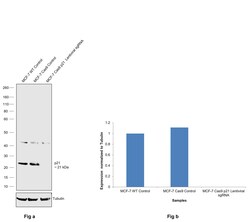
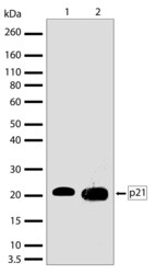
- CRISPR-Cas9 mediated genome editing ofp21 (as confirmed by next generation sequencing) was achieved by using LentiArray™ Lentiviral sgRNA (Product # A32042, AssayID CRISPR865095_LV) and LentiArray Cas9 Lentivirus (Product # A32064). Fig (a) Western blot analysis of p21 was performed by loading 30 µg of MCF-7 wild type (Lane 1), MCF-7 Cas9 (Lane 2) and MCF-7 Cas9 cells transduced with p21 Lentiviral sgRNA (Lane 3) whole cell extracts. The samples were electrophoresed using NuPAGE™ Novex™ 4-12% Bis-Tris Protein Gel (Product # NP0322BOX). Resolved proteins were then transferred onto a nitrocellulose membrane (Product # IB23001) by iBlot® 2 Dry Blotting System (Product # IB21001). The blot was probed with p21 Recombinant Rabbit Monoclonal Antibody (2H2L13) (Product # 701151, 1:1000 dilution) and Goat anti-Rabbit IgG (H+L) Superclonal™ Recombinant Secondary Antibody, HRP (Product # A27036, 1:20,000 dilution) using the iBright™ FL 1500 (Product # A44115). Chemiluminescent detection was performed using SuperSignal™ West Atto Ultimate Sensitivity Substrate (Product # A38556). A loss of signal in sgRNA transduced cells using the LentiArray™ CRISPR product line confirms that antibody is specific top21 (Fig (b)). Uncharacterized bands were observed at ~45 kDa in all the samples.
- Submitted by
- Invitrogen Antibodies (provider)
- Main image
- Experimental details
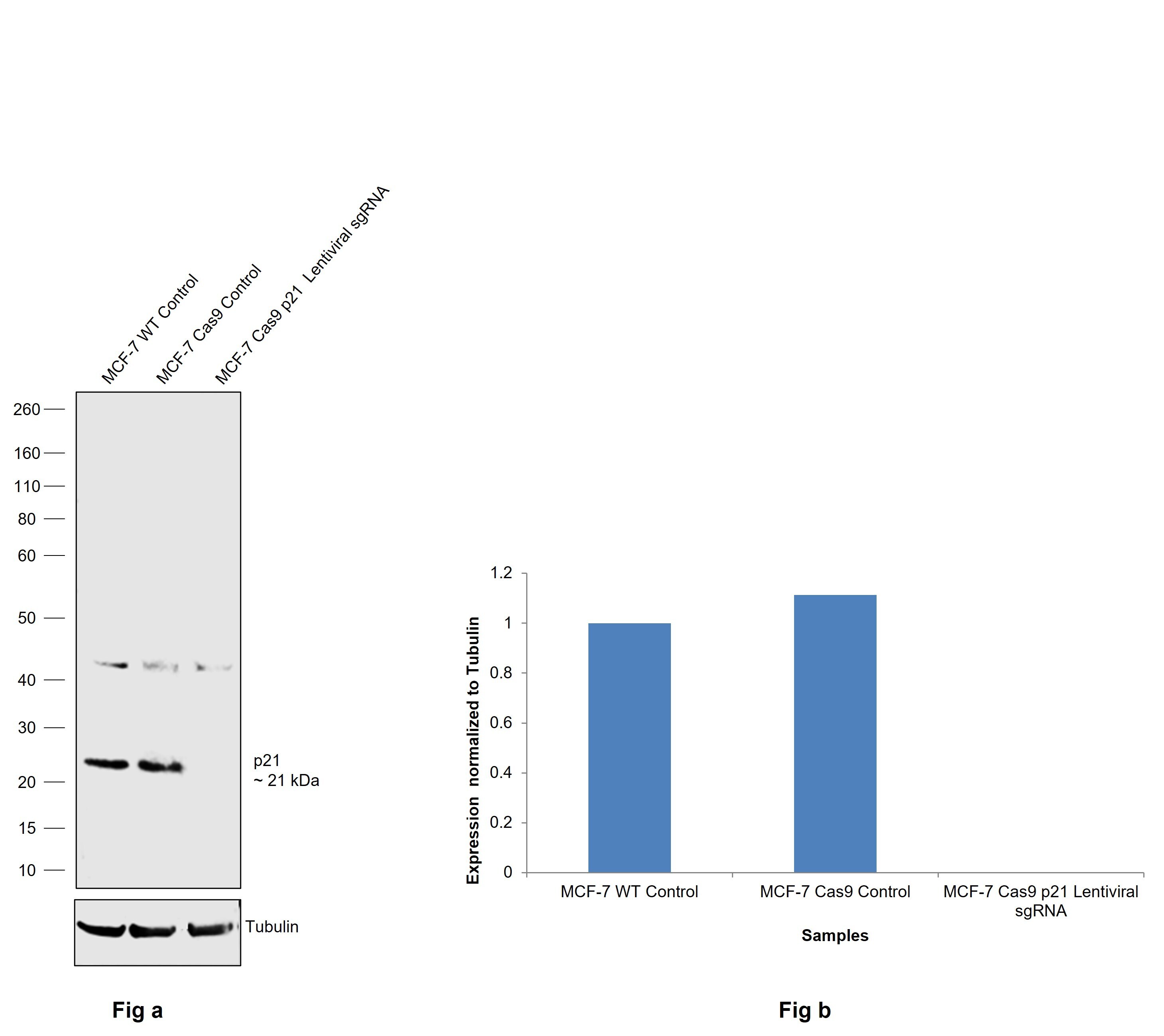
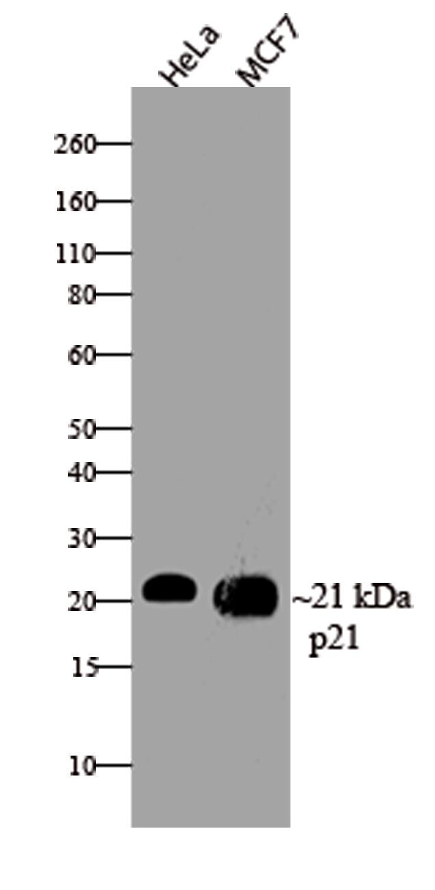
- Western blot analysis of p21 was performed by loading 30 µg of HeLa and MCF-7 cell lysates using Novex®NuPAGE®4-12% Bis-Tris gel (Product # NP0321BOX), XCell SureLock Electrophoresis System (Product # EI0002), Novex® Sharp Pre-Stained Protein Standard (Product # LC5800), and iBlot® Dry Blotting System (Product # IB21001). Proteins were transferred to a nitrocellulose membrane and blocked with 5% skim milk for 1 hour at room temperature. p21 was detected at ~21 kDa using p21 Recombinant Rabbit Monoclonal Antibody (Product # 701151) at a 1:1000 dilution in 2.5% skim milk at 4°C overnight on a rocking platform. Detection was performed using an HRP-conjugated Goat anti-Rabbit secondary antibody (Product # G-21234) at a 1:5000 dilution and chemiluminescent detection was performed using Pierce™ ECL Western blotting Substrate (Product # 32106).
- Submitted by
- Invitrogen Antibodies (provider)
- Main image
- Experimental details
- Western blot analysis of p21 in whole cell extracts of HeLa (lane 1) and MCF-7 (lane 2) using a p21 recombinant rabbit monoclonal antibody (Product # 701151) at a dilution of 2 µg/mL. Samples were detected using chemiluminescence (ECL). Results show a band at ~21kDa.

Supportive validation
- Submitted by
- Invitrogen Antibodies (provider)
- Main image
- Experimental details
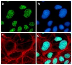
- Immunofluorescent analysis of p21 was performed on 70% confluent log phase U-2 OS cells. The cells were fixed with 4% paraformaldehyde for 15 minutes, permeabilized with 0. 25% Triton X-100 for 10 minutes, and blocked with 5% BSA for 1 hour at room temperature. The cells were labeled with p21 Recombinant Rabbit Monoclonal Antibody (Product # 701151) at a dilution of 1:500 in 1% BSA and incubated for 3 hours at room temperature and then labeled with Alexa Fluor® 488 Goat anti-Rabbit IgG secondary antibody (Product # A-11008) at a dilution of 1:400 for 30 minutes at room temperature (Panel a: green). Nuclei (Panel b: blue) were stained with SlowFade® Gold Antifade Mountant with DAPI (Product # S36938). F-actin (Panel c: red) was stained with Alexa Fluor® 594 phalloidin (Product # A12381). Panel d is a merged image showing nuclear localization. The images were captured using a Nikon microscope at 20X magnification.

Supportive validation
- Submitted by
- Invitrogen Antibodies (provider)
- Main image
- Experimental details
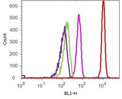
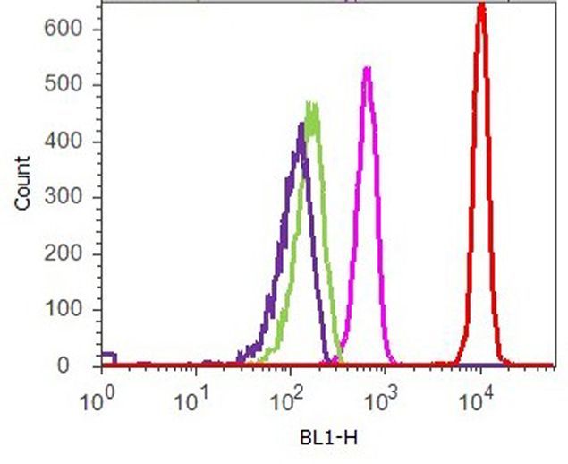
- Flow cytometry analysis of p21 was performed on serum-starved HeLa cells. Cells were fixed with 70% ethanol for 10 minutes, permeabilized with 0. 25% Tritonª X-100 for 20 minutes, and blocked with 5% BSA for 1 hour at room temperature. Cells were labeled with ABfinityª p21 recombinant rabbit monoclonal antibody (Product # 701151, red histogram) or with rabbit isotype control (pink histogram) at a dilution of 1:250 in 2.5% BSA. After incubation at room temperature for 3 hours, the cells were labeled with Alexa Fluor¨ 488 goat anti-Rabbit Secondary antibody (Product # A11008) at a dilution of 1:400 for 30 minutes at room temperature. The representative 10,000 cells were acquired and analyzed for each sample using an Attune¨ Acoustic Focusing Cytometer. The purple histogram represents unstained control cells and the green histogram represents no-primary-antibody control.
Supportive validation
- Submitted by
- Invitrogen Antibodies (provider)
- Main image

- Experimental details
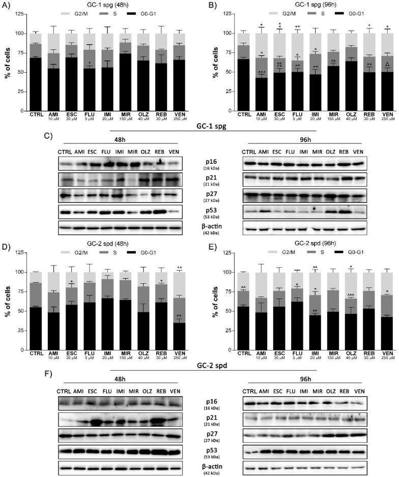
- Figure 2 Antidepressant-induced cell cycle profile regulation through related genes activation. GC-1 spg and GC-2 spd cells were treated with antidepressants for 48 and 96 h, the cell cycle profile ( A , B , D , E ) was determined and the level of p16, p21, p27, p53 proteins was controlled by Western blot technique. Representative blots are shown ( C , F ). Statistical differences were determined using one-way analysis of variance (ANOVA) with Dunnett's post-hoc test; p values < 0.05 were considered statistically significant. Asterisks (*) indicate the comparison between control and antidepressants-treated cells, whereas carets (^) indicate the comparison between the same drugs in different periods (48 vs. 96). Bars indicate mean +- SD, n = 3, ***/^^^ p < 0.001, **/^^ p < 0.01, */^ p < 0.05, no indication-no statistical significance.
- Submitted by
- Invitrogen Antibodies (provider)
- Main image
- Experimental details
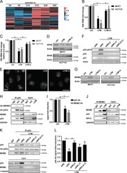
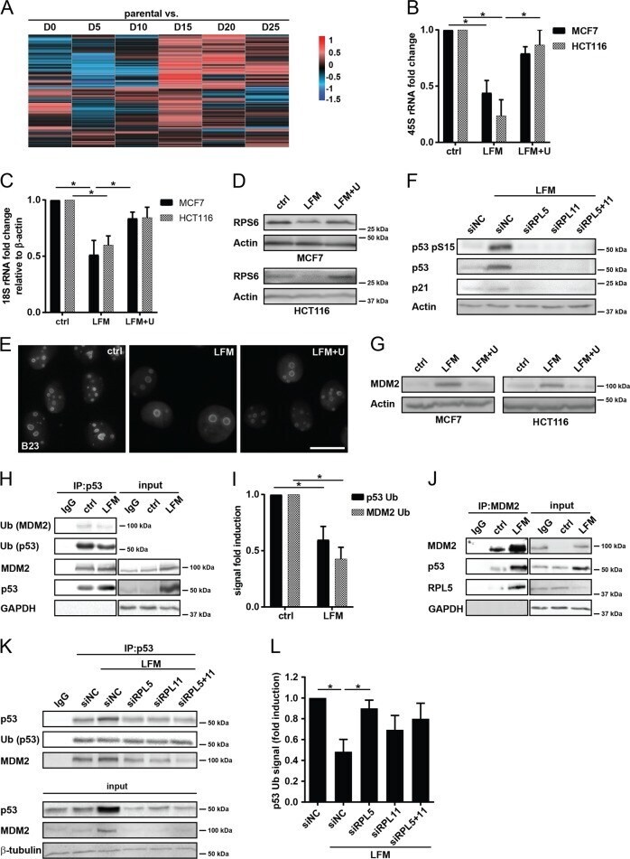
- Fig. 2 Inhibition of DHODH stabilizes the p53 tumor suppressor protein. a Parental 4T1 cells and D0-D25 sublines were subjected to microchip analysis and a heat map for the 119 transcripts involved in ribosome biogenesis was constructed. b - e , g MCF7 and HCT116 cells were exposed to leflunomide (LFM, 50 uM) for 72 h in the presence or absence of uridine (U; 50 ug/l). Expression of 45S rRNA ( b ) and 18S rRNA ( c ) was assessed by qRT-PCR. d Immunoblotting detection of RPS6 was performed. beta-Actin was used as a loading control. e Immunofluorescent detection of B23 in MCF7 cells was performed. f Immunoblotting detection of p53 pS15, p53, and p21 in MCF7 cells treated 72 h with LFM (50 uM) after downregulation of RPL5 and RPL11 alone or in combination using specific siRNA. Non-targeting siRNA (siNC) was used as a control. beta-Actin was used as a loading control. g Immunoblot detection of MDM2 in MCF7 and HCT116 cells. beta-Actin was used as a loading control. h Co-immunoprecipitation of p53 followed by immunoblot analysis of p53, MDM2, ubiquitinated p53, and ubiquitinated MDM2 in MCF7 control and LFM (50 uM, 72 h)-treated cells. GAPDH was used as an input control. Immunoprecipitated samples and input represent one membrane with different intensity of signal. i Levels of ubiquitinated p53 and MDM2 related to their immunoprecipitated total forms were evaluated from three independent experiments. j Co-immunoprecipitation of MDM2 followed by immunoblot analysi
- Submitted by
- Invitrogen Antibodies (provider)
- Main image

- Experimental details
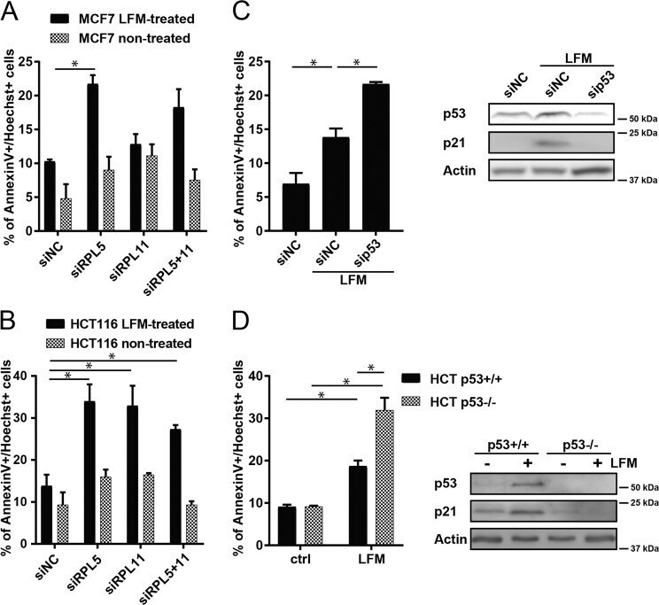
- Fig. 3 Role of p53 in cell cycle arrest after leflunomide treatment. a MCF7 and b HCT116 cells were exposed to LFM (50 uM) for 72 h after downregulation of RPL5 and RPL11 alone or in combination using specific siRNA. Cell death was assessed by flow cytometry using annexin V/Hoechst staining. Non-targeting siRNA (siNC) was used as a control. c MCF7 cells were transfected with specific p53 siRNA or with non-targeting siRNA (siNC), respectively. Cell death was evaluated by annexin V/Hoechst positivity using FACS. Immunoblot detection of p53 and p21 shows the effect of transfection. beta-Actin was used as a loading control. d HCT116 wt p53 and HCT116 p53 KO cells were exposed to LFM (50 uM) for 72 h and cell death was determined by flow cytometry using annexin V/Hoechst staining. Immunoblot detection of p53 and p21 show the effect of p53 KO . beta-Actin was used as a loading control. In a - d , data are shown as mean +- SEM, n = 3-5. * P < 0.05, two-way ANOVA.