 Explore
 Validate
 Learn
Learn Western blot
Antibody data
- Antibody Data
- Antigen structure
- References [1]
- Comments [0]
- Validations
- Western blot [4]
- Immunocytochemistry [1]
- Immunohistochemistry [1]
Submit
Validation data
Reference
Comment
Report error
- Product number
- PA5-36810 - Provider product page

- Provider
- Invitrogen Antibodies
- Product name
- Phospho-HDAC1 (Ser421) Polyclonal Antibody
- Antibody type
- Polyclonal
- Antigen
- Synthetic peptide
- Description
- This antibody detects endogenous protein at a molecular weight of 55 kDa.
- Concentration
- 1 mg/mL
Submitted references Deletion of Nemo-like Kinase in T Cells Reduces Single-Positive CD8(+) Thymocyte Population.
Daams R, Sime W, Leandersson K, Sitnicka E, Massoumi R
Journal of immunology (Baltimore, Md. : 1950) 2020 Oct 1;205(7):1830-1841
Journal of immunology (Baltimore, Md. : 1950) 2020 Oct 1;205(7):1830-1841
No comments: Submit comment
Supportive validation
- Submitted by
- Invitrogen Antibodies (provider)
- Main image

- Experimental details
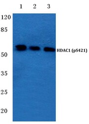
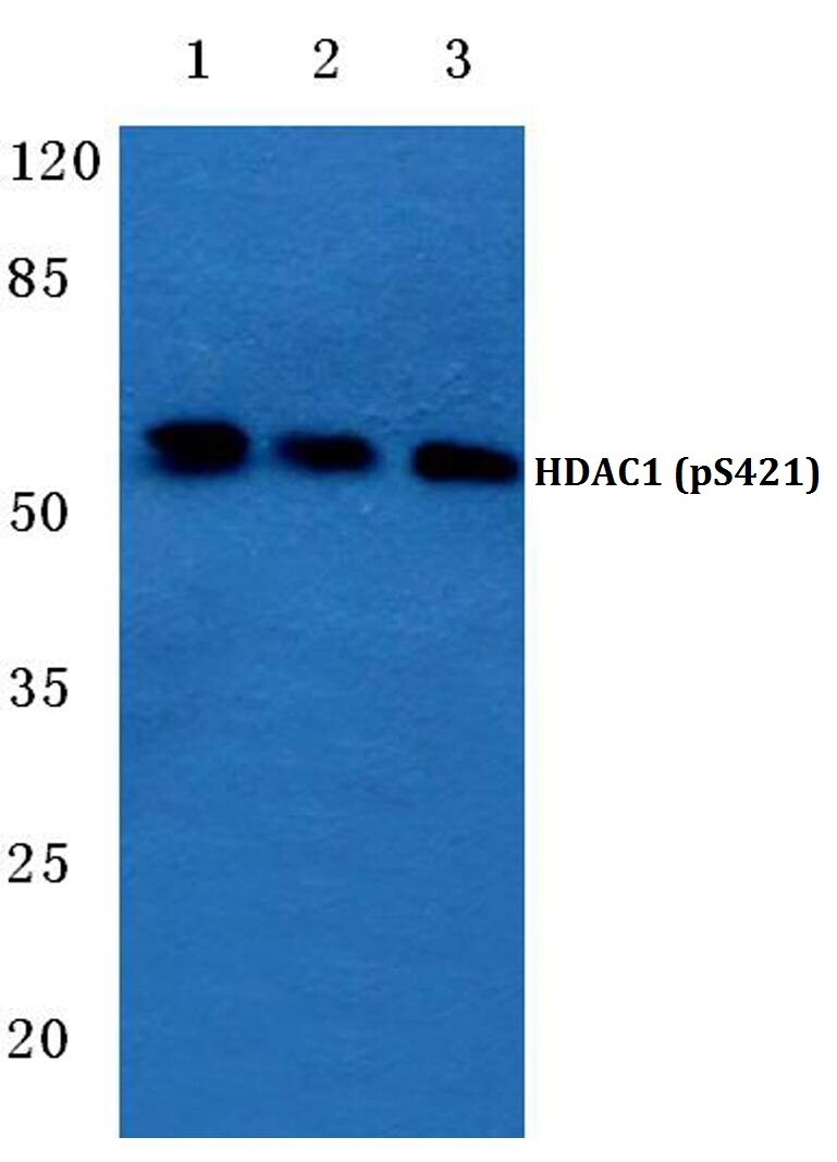
- Western blot analysis of Phospho-HDAC1 (Ser421) in Lane 1: MCF-7 cell lysate treated with EGF (0.1 ng/mL, 30 mins), Lane 2: mouse spleen tissue lysate, Lane 3: H9C2 cell lysate treated with EGF (0.1 ng/mL, 30 mins). Samples were incubated with Phospho-HDAC1 (Ser421) polyclonal antibody (Product # PA5-36810) at a dilution of 1:500.

- Submitted by
- Invitrogen Antibodies (provider)
- Main image

- Experimental details
- Western blot analysis of Phospho-HDAC1 pSer421 using Phospho-HDAC1 pSer421 polyclonal antibody (Product # PA5-36810) at a dilution of 1:500. Lane 1: MCF-7 cell lysate treated with EGF (0.1 ng/mL, 30 min), Lane 2: Mouse spleen tissue lysate, Lane 3: H9C2 cell lysate treated with EGF (0.1 ng/mL, 30 min).

- Submitted by
- Invitrogen Antibodies (provider)
- Main image

- Experimental details
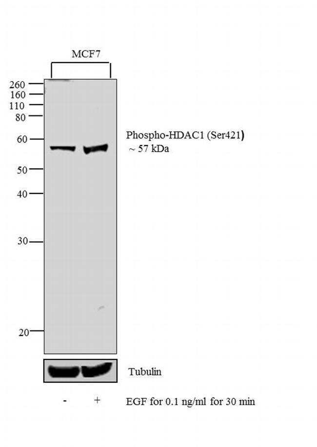
- Western blot analysis was performed on modified whole extracts (1% SDS) (30 µg lysate) of MCF7 (Lane 1) and MCF7 treated with EGF (0.1 ng/ml for 30 min) (Lane 2). The blot was probed with Anti-Phospho-HDAC1 (Ser421) Polyclonal Antibody (Product # PA5-36810, 1:1000 dilution) and detected by chemiluminescence using Goat anti-Rabbit IgG (H+L) Superclonal™ Secondary Antibody, HRP conjugate (Product # A27036, 0.25 µg/ml, 1:4000 dilution). A 57 kDa band corresponding to Phospho-HDAC1 (Ser421) was observed in the cell line tested and increased upon EGF treatment.
- Submitted by
- Invitrogen Antibodies (provider)
- Main image
- Experimental details
- Western blot analysis of Phospho-HDAC1 (Ser421) in Lane 1: MCF-7 cell lysate treated with EGF (0.1 ng/mL, 30 mins), Lane 2: mouse spleen tissue lysate, Lane 3: H9C2 cell lysate treated with EGF (0.1 ng/mL, 30 mins). Samples were incubated with Phospho-HDAC1 (Ser421) polyclonal antibody (Product # PA5-36810) at a dilution of 1:500.
Supportive validation
- Submitted by
- Invitrogen Antibodies (provider)
- Main image

- Experimental details
- Immunofluorescence analysis of Phospho-HDAC1 (Ser421) was performed using 70% confluent log phase MCF-7 cells treated with EGF (0.1 ng/mL for 30 min). The cells were fixed with 4% paraformaldehyde for 10 minutes, permeabilized with 0.1% Triton™ X-100 for 15 minutes, and blocked with 1% BSA for 1 hour at room temperature. The cells were labeled with Phospho-HDAC1 (Ser421) Polyclonal Antibody (Product # PA5-36810) at 5 µg/mL in 0.1% BSA, incubated at 4 degree celsius overnight and then labeled with Goat anti-Rabbit IgG (H+L) Superclonal™ Secondary Antibody, Alexa Fluor® 488 conjugate (Product # A27034) at a dilution of 1:2000 for 45 minutes at room temperature (Panel a: green). Nuclei (Panel b: blue) were stained with ProLong™ Diamond Antifade Mountant with DAPI (Product # P36962). F-actin (Panel c: red) was stained with Rhodamine Phalloidin (Product # R415, 1:300). Panel d represents the merged image showing nuclear signal upon EGF treatment. Panel e represents the control cells showing very weak staining in the nucleus. Panel f represents control cells with no primary antibody to assess background. The images were captured at 60X magnification.

Supportive validation
- Submitted by
- Invitrogen Antibodies (provider)
- Main image
- Experimental details
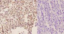
- Immunohistochemistry analysis of Phospho-HDAC1 (Ser421) in paraffin-embedded human tonsil carcinoma tissue (nucleus staining) and negative control (right, with PBS only). Samples were incubated with Phospho-HDAC1 (Ser421) polyclonal antibody (Product # PA5-36810) at a dilution of 1:50, followed by goat Anti-Rabbit IgG-biotin and avidin peroxidase.
