 Explore
 Validate
 Learn
Learn Western blot
 ELISA
ELISAAntibody data
- Antibody Data
- Antigen structure
- References [0]
- Comments [0]
- Validations
- Western blot [2]
Submit
Validation data
Reference
Comment
Report error
- Product number
- GTX23707 - Provider product page

- Provider
- GeneTex
- Proper citation
- GeneTex Cat#GTX23707, RRID:AB_423842
- Product name
- Ap2A antibody
- Antibody type
- Polyclonal
- Reactivity
- Human, Mouse, Rat, Canine, Chicken/Avian
- Host
- Goat
- Storage
- Store vial at -20°C prior to opening. Aliquot contents and freeze at -20°C or below for extended storage. Avoid cycles of freezing and thawing.
No comments: Submit comment
Supportive validation
- Submitted by
- GeneTex (provider)
- Main image

- Experimental details
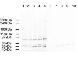
- Western blot using GeneTex's Affinity Purified anti-AP2A antibody shows detection of a band just below 100 kDa correspond-ing to Human AP2A1 in a various preparations. Lane 1 - HeLa nuclear extract, Lane 2 - HeLa, Lane 3 - 293, Lane 4 - A431 and Lane 5 - Jurkat whole cell lysates. In lanes 6-10 the antibody was preincubated with 1 µg/ml of the immunizing peptide which effect-ively blocks the specific reactivity of this antibody with AP2A. Approximately 20 µg of each lysate was run on a SDS-PAGE and transferred onto nitrocellulose followed by reaction with a 1:500 dilution of anti-AP2A antibody. Detection occurred using a 1:5,000 dilution of HRP-labeled Rabbit anti-Goat IgG for 1 hour at room temperature. A chemi-luminescence system was used for signal detection (Roche) using a 60-sec exposure time.
- Validation comment
- WB

- Submitted by
- GeneTex (provider)
- Main image
- Experimental details
- Western blot using GeneTex's Affinity Purified anti-AP2A antibody shows detection of a band just below 100 kDa correspond-ing to Human AP2A1 in a various preparations. Lane 1 - HeLa nuclear extract, Lane 2 - HeLa, Lane 3 - 293, Lane 4 - A431 and Lane 5 - Jurkat whole cell lysates. In lanes 6-10 the antibody was preincubated with 1 ?g/ml of the immunizing peptide which effect-ively blocks the specific reactivity of this antibody with AP2A. Approximately 20 ?g of each lysate was run on a SDS-PAGE and transferred onto nitrocellulose followed by reaction with a 1:500 dilution of anti-AP2A antibody. Detection occurred using a 1:5,000 dilution of HRP-labeled Rabbit anti-Goat IgG for 1 hour at room temperature. A chemi-luminescence system was used for signal detection (Roche) using a 60-sec exposure time.
