Explore
 Validate
 Learn
Learn Western blot
Antibody data
- Antibody Data
- Antigen structure
- References [1]
- Comments [0]
- Validations
- Western blot [1]
- Immunocytochemistry [3]
- Immunohistochemistry [1]
- Flow cytometry [1]
- Other assay [2]
Submit
Validation data
Reference
Comment
Report error
- Product number
- MA5-32078 - Provider product page

- Provider
- Invitrogen Antibodies
- Product name
- CD86 (B7-2) Recombinant Rabbit Monoclonal Antibody (SJ20-00)
- Antibody type
- Monoclonal
- Antigen
- Synthetic peptide
- Description
- Recombinant rabbit monoclonal antibodies are produced using in vitro expression systems. The expression systems are developed by cloning in the specific antibody DNA sequences from immunoreactive rabbits. Then, individual clones are screened to select the best candidates for production. The advantages of using recombinant rabbit monoclonal antibodies include: better specificity and sensitivity, lot-to-lot consistency, animal origin-free formulations, and broader immunoreactivity to diverse targets due to larger rabbit immune repertoire.
- Reactivity
- Human, Rat
- Host
- Rabbit
- Isotype
- IgG
- Antibody clone number
- SJ20-00
- Vial size
- 100 µL
- Concentration
- 1 mg/mL
- Storage
- Store at 4°C short term. For long term storage, store at -20°C, avoiding freeze/thaw cycles.
Submitted references Urinary Trypsin Inhibitor Protects Tight Junctions of Septic Pulmonary Capillary Endothelial Cells by Regulating the Functions of Macrophages.
Wang R, Song W, Xie C, Zhong W, Xu H, Zhou Q, Deng Y, Hong Y, Li X, Fang M
Journal of inflammation research 2021;14:1973-1989
Journal of inflammation research 2021;14:1973-1989
No comments: Submit comment
Supportive validation
- Submitted by
- Invitrogen Antibodies (provider)
- Main image
- Experimental details
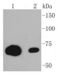
- Western blot analysis of CD86 (B7-2) in different lysates using a Monoclonal antibody (Product #MA5-32078) at a dilution of 1:1,000. Positive control: Lane 1: Raji, Lane 2: Jurkat.

Supportive validation
- Submitted by
- Invitrogen Antibodies (provider)
- Main image

- Experimental details
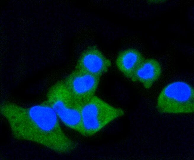
- Immunocytochemical analysis of CD86 (B7-2) in HUVEC cells using a CD86 (B7-2) Monoclonal antibody (Product # MA5-32078) as seen in green. The nuclear counter stain is DAPI (blue). Cells were fixed in paraformaldehyde, permeabilised with 0.25% Triton X100/PBS.

- Submitted by
- Invitrogen Antibodies (provider)
- Main image

- Experimental details
- Immunocytochemical analysis of CD86 (B7-2) in JAR cells using a CD86 (B7-2) Monoclonal antibody (Product # MA5-32078) as seen in green. The nuclear counter stain is DAPI (blue). Cells were fixed in paraformaldehyde, permeabilised with 0.25% Triton X100/PBS.

- Submitted by
- Invitrogen Antibodies (provider)
- Main image

- Experimental details
- Immunocytochemical analysis of CD86 (B7-2) in Hela cells using a CD86 (B7-2) Monoclonal antibody (Product # MA5-32078) as seen in green. The nuclear counter stain is DAPI (blue). Cells were fixed in paraformaldehyde, permeabilised with 0.25% Triton X100/PBS.
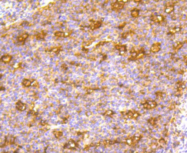
Supportive validation
- Submitted by
- Invitrogen Antibodies (provider)
- Main image
- Experimental details
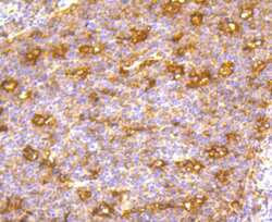
- Immunohistochemical analysis of CD86 (B7-2) of paraffin-embedded Human tonsil tissue using a CD86-B7-2 Monoclonal antibody (Product #MA5-32078). Counter stained with hematoxylin.
Supportive validation
- Submitted by
- Invitrogen Antibodies (provider)
- Main image
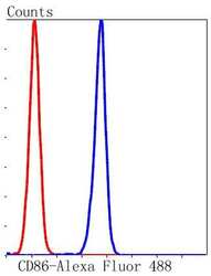
- Experimental details
- Flow Cytometric analysis of CD86 (B7-2) in K562 cells using a CD86 (B7-2) Monoclonal Antibody (Product # MA5-32078) at a dilution of 1:50, as seen in blue compared with an unlabelled control (cells without incubation with primary antibody; red). Alexa Fluor 488-conjugated goat anti rabbit IgG was used as the secondary antibody.

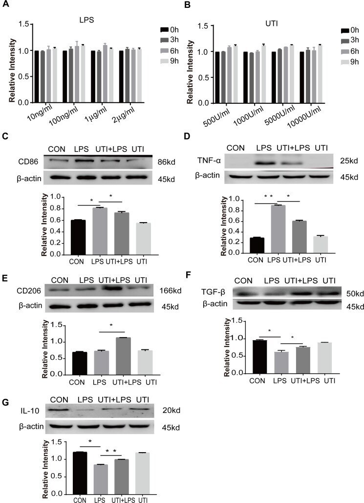
Supportive validation
- Submitted by
- Invitrogen Antibodies (provider)
- Main image

- Experimental details
- Figure 1 Effects of UTI and LPS on viability of BMDM. In the range of 10-2000 ng/mL, LPS ( A ) did not affect the cell viability at 0-9 h. UTI ( B ) at the concentration between 500 U/mL and 10 4 U/mL did not affect the viability of cells at 0-9 h. UTI regulated polarization of M1 to M2 macrophage when incubated with LPS. Western blot showing expression level of CD86 ( C ) and TNF-alpha ( D ) that was markedly increased in the LPS group when compared with the control group; it was significantly decreased when pretreated with UTI. CD206 ( E ), TGF-beta ( F ) and IL-10 ( G ) expression was markedly decreased in the LPS group in comparison with the control group; it was augmented when pretreated with UTI. * p
- Submitted by
- Invitrogen Antibodies (provider)
- Main image
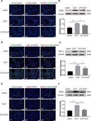
- Experimental details
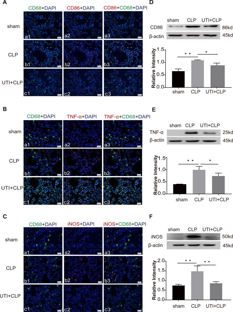
- Figure 5 UTI decreased M1 macrophages in lung tissue in CLP-induced rat lungs. CD86 ( A b1-3), TNF-alpha ( B b1-3), iNOS ( C b1-3) expression (red) was intensely labeled in lung tissue of the CLP group. The expression decreased in the UTI+CLP group ( A c1-3, B c1-3, C c1-3). It was hardly detected in the sham group (( A a1-3, B a1-3, C a1-3). Western blot showing the expression of CD86 ( D ), TNF-alpha ( E ) and iNOS ( F ) sharply increased in the CLP group in comparison with the sham group; it significantly decreased in the UTI+CLP group. * p