 Explore
 Validate
 Learn
Learn Western blot
Antibody data
- Antibody Data
- Antigen structure
- References [0]
- Comments [0]
- Validations
- Western blot [3]
- Immunocytochemistry [1]
Submit
Validation data
Reference
Comment
Report error
- Product number
- MA5-15386 - Provider product page

- Provider
- Invitrogen Antibodies
- Product name
- YES1 Monoclonal Antibody (2F3E6)
- Antibody type
- Monoclonal
- Antigen
- Purifed from natural sources
- Description
- MA5-15386 targets YES1 in WB applications and shows reactivity with Human samples.
- Antibody clone number
- 2F3E6
- Concentration
- Conc. Not Determined
No comments: Submit comment
Supportive validation
- Submitted by
- Invitrogen Antibodies (provider)
- Main image
- Experimental details
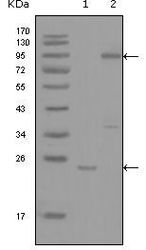
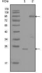
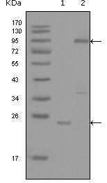
- Western blot analysis of YES1 using a YES1 monoclonal antibody (Product # MA5-15386) against a truncated YES1-His recombinant protein (1) and full-length GFP-YES1 (aa1-543) transfected COS-7 cell lysate (2).
- Submitted by
- Invitrogen Antibodies (provider)
- Main image
- Experimental details
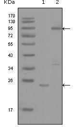
- Western blot analysis of YES1 using a YES1 monoclonal antibody (Product # MA5-15386) against a truncated YES1-His recombinant protein (1) and full-length GFP-YES1 (aa1-543) transfected COS-7 cell lysate (2).
- Submitted by
- Invitrogen Antibodies (provider)
- Main image
- Experimental details
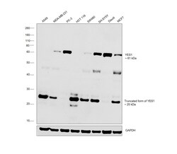
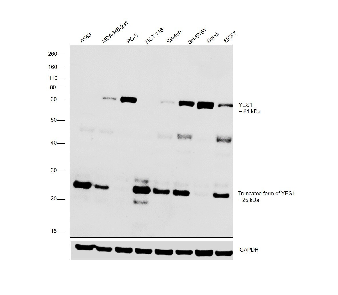
- Western blot was performed using Anti-YES1 Monoclonal Antibody (2F3E6) (Product # MA5-15386) and a band at 61 kDa corresponding to full length YES1, ~ 25 kDa band corresponding to truncated form of YES1 were observed across the cell lines tested. Whole cell extracts (30 µg lysate) of A549 (Lane 1), MDA-MB-231 (Lane 2), PC-3 (Lane 3), HCT 116 (Lane 4), SW480 (Lane 5), SH-SY5Y (Lane 6), Daudi (Lane 7) MCF7 (Lane 8) were electrophoresed using Novex® NuPAGE® 4-12% % Bis-Tris gel (Product # NP0321BOX). Resolved proteins were then transferred onto a nitrocellulose membrane (Product # IB23001) by iBlot® 2 Dry Blotting System (Product # IB21001). The blot was probed with the primary antibody (1:500 dilution) and detected by chemiluminescence with Goat anti-Mouse IgG (H+L) Superclonal™ Recombinant Secondary Antibody, HRP (Product # A28177, 1:4000 dilution) using the iBright FL 1000 (Product # A32752). Chemiluminescent detection was performed using Novex® ECL Chemiluminescent Substrate Reagent Kit (Product # WP20005).
Supportive validation
- Submitted by
- Invitrogen Antibodies (provider)
- Main image
- Experimental details
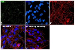
- Immunofluorescence analysis of YES1 was performed using 70% confluent log phase SH-SY5Y cells. The cells were fixed with 4% paraformaldehyde for 10 minutes, permeabilized with 0.1% Triton™ X-100 for 15 minutes, and blocked with 2% BSA for 1 hour at room temperature. The cells were labeled with YES1 Monoclonal Antibody (2F3E6) (Product # MA5-15386) at 1:100 dilution in 0.1% BSA, incubated at 4 degree Celsius overnight and then labeled with Goat anti-Mouse IgG (H+L) Highly Cross-Adsorbed Secondary Antibody, Alexa Fluor Plus 488 (Product # A32723) at a dilution of 1:2000 for 45 minutes at room temperature (Panel a: green). Nuclei (Panel b: blue) were stained with ProLong™ Diamond Antifade Mountant with DAPI (Product # P36962). F-actin (Panel c: red) was stained with Rhodamine Phalloidin (Product # R415, 1:300). Panel d represents the merged image showing cytoplasmic localization. Panel e represents control cells with no primary antibody to assess background. The images were captured at 60X magnification.
