Explore
 Validate
 Learn
LearnPA5-14253
antibody from Invitrogen Antibodies
Targeting: SERPINH1
CBP1, CBP2, colligen, HSP47, SERPINH2
 Western blot
Antibody data
- Antibody Data
- Antigen structure
- References [0]
- Comments [0]
- Validations
- Western blot [3]
- Immunohistochemistry [1]
- Flow cytometry [1]
Submit
Validation data
Reference
Comment
Report error
- Product number
- PA5-14253 - Provider product page

- Provider
- Invitrogen Antibodies
- Product name
- SERPINH1 Polyclonal Antibody
- Antibody type
- Polyclonal
- Antigen
- Synthetic peptide
- Description
- This antibody is predicted to react with bovine, chicken, mouse and rat based on sequence homology.
- Reactivity
- Human
- Host
- Rabbit
- Isotype
- IgG
- Vial size
- 400 µL
- Concentration
- 2 mg/mL
- Storage
- -20° C, Avoid Freeze/Thaw Cycles
No comments: Submit comment
Supportive validation
- Submitted by
- Invitrogen Antibodies (provider)
- Main image
- Experimental details
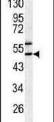
- Western blot analysis using a SERPINH1 polyclonal antibody (Product # PA5-14253) in HeLa cell lysates (35 µg per lane).

- Submitted by
- Invitrogen Antibodies (provider)
- Main image

- Experimental details
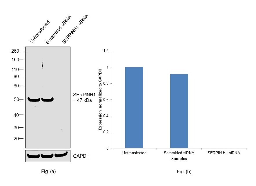
- Knockdown of Serpin H1 was achieved by transfecting Caco-2 with Serpin H1 specific siRNAs (Silencer® select Product # s2485, s2486). Western Blot analysis (Fig. a) was performed using Whole cell extracts from the Serpin H1 knockdown cells (lane 3), non-targeting scrambled siRNA transfected cells (lane 2) and untransfected cells (lane 1). The blot was probed with SERPINH1 Polyclonal Antibody (Product # PA5-14253, 1:1000 dilution ) and Goat anti-Rabbit IgG (H+L) Superclonal™ Recombinant Secondary Antibody, HRP (Product # A27036, 1:20000 dilution). Densitometric analysis of this Western Blot is shown in histogram (Fig. b). Decrease in signal upon siRNA mediated knock down confirms that antibody is specific to Serpin H1.
- Submitted by
- Invitrogen Antibodies (provider)
- Main image
- Experimental details
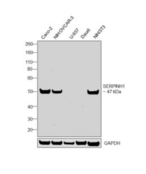
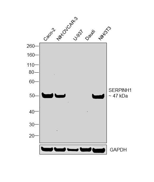
- Western Blot was performed using Anti-SERPINH1 Polyclonal Antibody (Product # PA5-14253) and a 47 kDa band corresponding to Serpin H1 was observed across tested cell lines. Whole cell extracts (40 µg lysate) of Caco-2 (Lane 1), NIH:OVCAR-3 (Lane 2), U-937 (Lane 3), Daudi (Lane 4), NIH/3T3 (Lane 5) were electrophoresed using NuPAGE™ 4-12% Bis-Tris Protein Gel (Product # NP0321BOX). Resolved proteins were then transferred onto a nitrocellulose membrane (Product # IB23001) by iBlot® 2 Dry Blotting System (Product # IB21001). The blot was probed with the primary antibody (1:1000 dilution) and detected by chemiluminescence with Goat anti-Rabbit IgG (H+L) Superclonal™ Recombinant Secondary Antibody, HRP (Product # A27036, 1:20000 dilution) using the iBright FL 1000 (Product # A32752). Chemiluminescent detection was performed using SuperSignal™ West Dura Extended Duration Substrate (Product # 34076).
Supportive validation
- Submitted by
- Invitrogen Antibodies (provider)
- Main image
- Experimental details
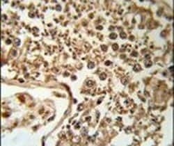
- Immunohistochemistry analysis in formalin-fixed, paraffin-embedded human testis tissue using a SERPINH1 polyclonal antibody (Product # PA5-14253), followed by HRP-conjugated secondary antibody and DAB staining.

Supportive validation
- Submitted by
- Invitrogen Antibodies (provider)
- Main image

- Experimental details
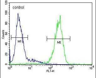
- Flow cytometry analysis of HepG2 cells using a SERPINH1 polyclonal antibody (Product # PA5-14253) (right) compared to a negative control cell (left) at a dilution of 1:10-50, followed by a FITC-conjugated goat anti-rabbit antibody