Explore
 Validate
 Learn
Learn Western blot
Antibody data
- Antibody Data
- Antigen structure
- References [0]
- Comments [0]
- Validations
- Western blot [2]
- Immunocytochemistry [1]
Submit
Validation data
Reference
Comment
Report error
- Product number
- MAB1205 - Provider product page

- Provider
- R&D Systems
- Product name
- Human/Mouse/Rat Phospho-JNK (T183/Y185) Antibody
- Antibody type
- Monoclonal
- Description
- Protein A or G purified from cell culture supernatant. Detects human, mouse and rat p46 and p54 JNK when dually phosphorylated at sites homologous to T183/Y185 of JNK1 and JNK2, and T221/Y223 of JNK3 in Western blots.
- Reactivity
- Human, Mouse, Rat
- Host
- Rabbit
- Conjugate
- Unconjugated
- Isotype
- IgG
- Antibody clone number
- 1006A
- Vial size
- 100 ug
- Storage
- Use a manual defrost freezer and avoid repeated freeze-thaw cycles. 12 months from date of receipt, -20 to -70 °C as supplied. 1 month, 2 to 8 °C under sterile conditions after reconstitution. 6 months, -20 to -70 °C under sterile conditions after reconstitution.
No comments: Submit comment
Supportive validation
- Submitted by
- R&D Systems (provider)
- Main image
- Experimental details
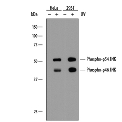
- Detection of Human and Mouse Phospho-JNK (T183/Y185) by Western Blot. Western blot shows lysates of HeLa human cervical epithelial carcinoma cell line and 293T human embryonic kidney cell line untreated (-) or treated (+) with 20 mJ/cm2 ultraviolet light (UV) followed by a 30 minute recovery. PVDF membrane was probed with 1 μg/ml of Rabbit Anti-Human/Mouse/Rat Phospho-JNK (T183/Y185) Monoclonal Antibody (Catalog # MAB1205), followed by HRP-conjugated Anti-Rabbit IgG Secondary Antibody (Catalog # HAF008). Specific bands were detected for Phospho-JNK (T183/Y185) at approximately 46 and 54 kDa (as indicated). This experiment was conducted under reducing conditions and using Immunoblot Buffer Group 1.
- Submitted by
- R&D Systems (provider)
- Main image

- Experimental details
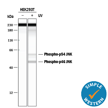
- Detection of Human Phospho-JNK (T183/Y185) by Simple WesternTM. Simple Western lane view shows lysates of HEK293T human embryonic kidney cell line untreated (-) or treated (+) with 20 J/m2 ultraviolet light (UV) followed by a 30 minute recovery, loaded at 0.2 mg/mL. A specific band was detected for Phospho-JNK (T183/Y185) at approximately 46 and 56 kDa (as indicated) using 20 μg/ml of Rabbit Anti-Human/Mouse/Rat Phospho-JNK (T183/Y185) Monoclonal Antibody (Catalog # MAB1205). This experiment was conducted under reducing conditions and using the 12-230 kDa separation system. Non-specific interaction with the 230 kDa Simple Western standard may be seen with this antibody.
Supportive validation
- Submitted by
- R&D Systems (provider)
- Main image

- Experimental details
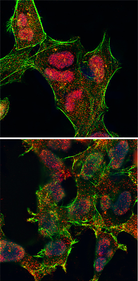
- Phospho-JNK (T183/Y185) in HEK293 Human Cell Line. JNK phosphorylated at T183/Y185 was detected in immersion fixed HEK293 human embryonic kidney cell line untreated (lower panel) or treated with UV radiation (upper panel) using Rabbit Anti-Human/Mouse/Rat Phospho-JNK (T183/Y185) Monoclonal Antibody (Catalog # MAB1205) at 25 μg/ml for 3 hours at room temperature. Cells were stained using the NorthernLights™ 557-conjugated Anti-Rabbit IgG Secondary Antibody (red; Catalog # NL004) and counterstained with DAPI. Filamentous actin was stained with fluorescein-conjugated phalloidin (green). Specific staining was localized to nuclei. View our protocol for Fluorescent ICC Staining of Cells on Coverslips.