 Explore
 Validate
 Learn
Learn Western blot
Antibody data
- Antibody Data
- Antigen structure
- References [0]
- Comments [0]
- Validations
- Western blot [4]
- Immunohistochemistry [7]
Submit
Validation data
Reference
Comment
Report error
- Product number
- LS-C344078 - Provider product page

- Provider
- LSBio
- Product name
- JUNB / JUN-B Antibody (aa323-347) LS-C344078
- Antibody type
- Polyclonal
- Description
- Immunogen affinity purified
- Reactivity
- Human, Mouse, Rat, Guinea Pig, Horse, Porcine, Simian
- Host
- Rabbit
- Storage
- At -20°C for 1 year. After reconstitution, at 4°C for 1 month. It can also be aliquotted and stored frozen at -20°C for a longer time. Avoid freeze-thaw cycles.
No comments: Submit comment
Supportive validation
- Submitted by
- LSBio (provider)
- Enhanced method
- Genetic validation
- Main image

- Experimental details
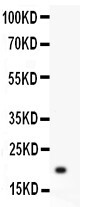
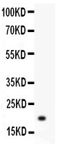
- JunB antibody Western blot. All lanes: Anti JunB at 0.5 ug/ml. WB: Recombinant Human JunB Protein 0.5ng. Predicted band size: 19 kD. Observed band size: 19 kD.
- Submitted by
- LSBio (provider)
- Enhanced method
- Genetic validation
- Main image

- Experimental details
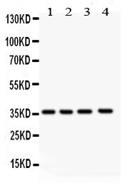
- JunB antibody Western blot. All lanes: Anti JunB at 0.5 ug/ml. Lane 1: Human Placenta Tissue Lysate at 50 ug. Lane 2: Rat Spleen Tissue Lysate at 50 ug. Lane 3: JURKAT Whole Cell Lysate at 40 ug. Lane 4: HELA Whole Cell Lysate at 40 ug. Predicted band size: 36 kD. Observed band size: 36 kD.
- Submitted by
- LSBio (provider)
- Main image

- Experimental details
- JunB antibody Western blot. All lanes: Anti JunB at 0.5 ug/ml. WB: Recombinant Human JunB Protein 0.5ng. Predicted band size: 19 kD. Observed band size: 19 kD.
- Submitted by
- LSBio (provider)
- Main image

- Experimental details
- JunB antibody Western blot. All lanes: Anti JunB at 0.5 ug/ml. Lane 1: Human Placenta Tissue Lysate at 50 ug. Lane 2: Rat Spleen Tissue Lysate at 50 ug. Lane 3: JURKAT Whole Cell Lysate at 40 ug. Lane 4: HELA Whole Cell Lysate at 40 ug. Predicted band size: 36 kD. Observed band size: 36 kD.

Supportive validation
- Submitted by
- LSBio (provider)
- Enhanced method
- Genetic validation
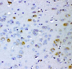
- Main image

- Experimental details
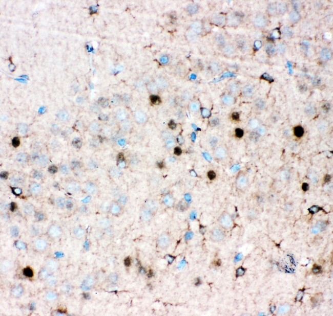
- JunB antibody IHC-paraffin: Mouse Brain Tissue.
- Submitted by
- LSBio (provider)
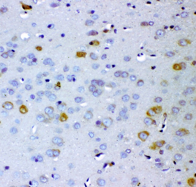
- Enhanced method
- Genetic validation
- Main image
- Experimental details
- JunB antibody IHC-paraffin: Rat Brain Tissue.
- Submitted by
- LSBio (provider)
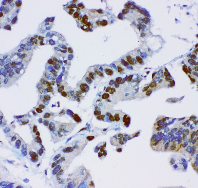
- Enhanced method
- Genetic validation
- Main image

- Experimental details
- JunB antibody IHC-paraffin: Human Intestinal Cancer Tissue.

- Submitted by
- LSBio (provider)
- Main image

- Experimental details
- JunB antibody IHC-paraffin: Mouse Brain Tissue.

- Submitted by
- LSBio (provider)
- Main image

- Experimental details
- JunB antibody IHC-paraffin: Rat Brain Tissue.

- Submitted by
- LSBio (provider)
- Main image

- Experimental details
- JunB antibody IHC-paraffin: Human Intestinal Cancer Tissue.
- Submitted by
- LSBio (provider)
- Main image

- Experimental details
- JunB antibody IHC-paraffin: Human Intestinal Cancer Tissue.
