Explore
 Validate
 Learn
Learn Western blot
Antibody data
- Antibody Data
- Antigen structure
- References [0]
- Comments [0]
- Validations
- Western blot [6]
- Immunocytochemistry [3]
- Immunohistochemistry [1]
- Chromatin Immunoprecipitation [1]
- Other assay [2]
Submit
Validation data
Reference
Comment
Report error
- Product number
- PA5-34758 - Provider product page

- Provider
- Invitrogen Antibodies
- Product name
- Ku80 Polyclonal Antibody
- Antibody type
- Polyclonal
- Antigen
- Recombinant protein fragment
- Description
- Recommended positive controls: 293T, A431, HepG2, A375, Neuro2A, PC-12, Rat2.
- Concentration
- 0.41 mg/mL
No comments: Submit comment
Supportive validation
- Submitted by
- Invitrogen Antibodies (provider)
- Main image
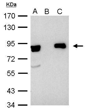
- Experimental details
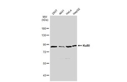
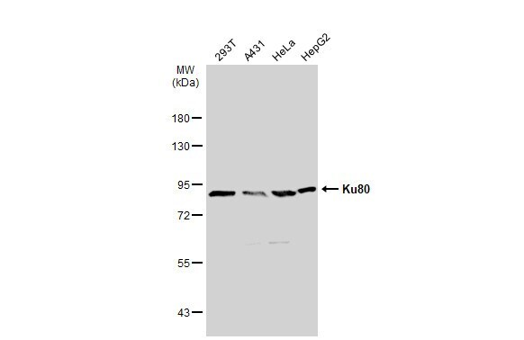
- Western blot analysis of Ku80 using A) 30 µg 293T whole cell lysate (B) 30 µg A431 whole cell lysate (C) 30 µg HeLa whole cell lysate and D) 30 µg HepG2 whole cell lysate. Samples were loaded onto a 7.5% SDS-PAGE gel and probed with a Ku80 polyclonal antibody (Product # PA5-34758) at a dilution of 1:1000.
- Submitted by
- Invitrogen Antibodies (provider)
- Main image

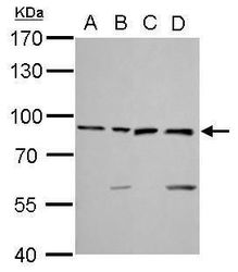
- Experimental details
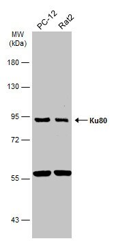
- Western Blot analysis of Ku80 was performed by separating 30 µg of Whole cell extracts by 7.5% SDS-PAGE. Proteins were transferred to a membrane and probed with a Ku80 Polyclonal Antibody (Product # PA5-34758) at a dilution of 1:1000. The HRP-conjugated anti-rabbit IgG antibody was used to detect the primary antibody.

- Submitted by
- Invitrogen Antibodies (provider)
- Main image
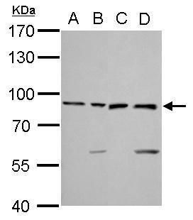
- Experimental details
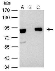
- Western Blot analysis of Ku80 was performed by separating 30 µg of various whole cell extracts by 7.5% SDS-PAGE. Proteins were transferred to a membrane and probed with a Ku80 Polyclonal Antibody (Product # PA5-34758) at a dilution of 1:1000 and a HRP-conjugated anti-rabbit IgG secondary antibody.
- Submitted by
- Invitrogen Antibodies (provider)
- Main image
- Experimental details
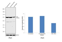
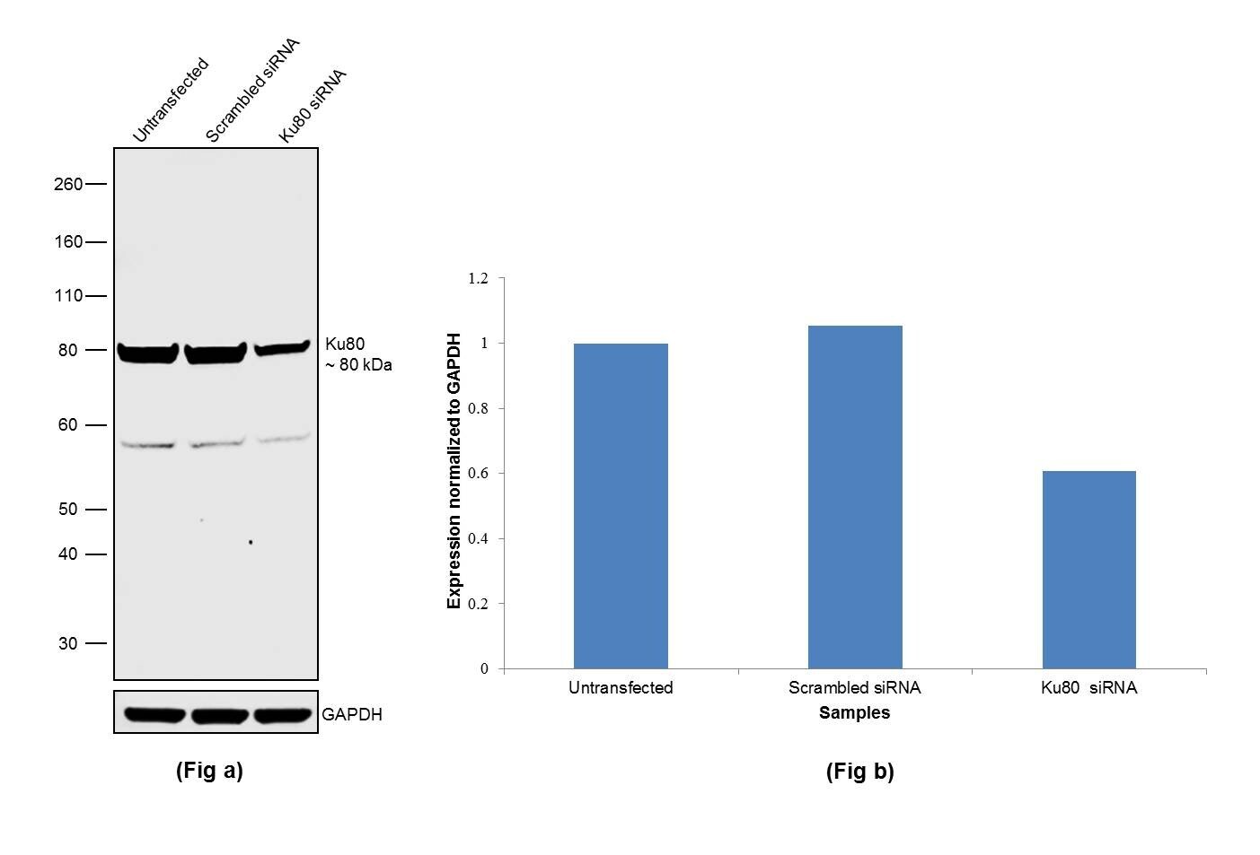
- Knockdown of Ku80 was achieved by transfecting HeLa with Ku80 specific siRNAs (Silencer® select Product # s14952, s14953). Western blot analysis (Fig. a) was performed using whole cell extracts from the Ku80 knockdown cells (lane 3), non-specific scrambled siRNA transfected cells (lane 2) and untransfected cells (lane 1). The blot was probed with Ku80 Polyclonal Antibody (Product # PA5-34758, 1:1000 dilution) and Goat anti-Rabbit IgG (H+L) Superclonal™ Recombinant Secondary Antibody, HRP (Product # A27036, 0.25µg/ml, 1:4000 dilution). Densitometric analysis of this western blot is shown in histogram (Fig. b). Decrease in signal upon siRNA mediated knock down confirms that antibody is specific to Ku80.
- Submitted by
- Invitrogen Antibodies (provider)
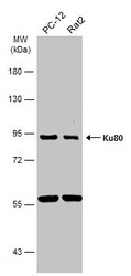
- Main image

- Experimental details
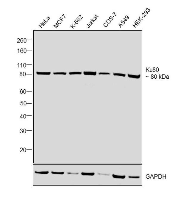
- Western blot was performed using Anti-Ku80 Polyclonal Antibody, (Product # PA5-34758) and a 80 kDa band corresponding to Ku80 was observed in the cell lines tested. Modified whole cell extracts (1%SDS) (30 µg lysate) of HeLa (Lane 1), MCF7 (Lane 2), K-562 (Lane 3), Jurkat (Lane 4), COS-7 (Lane 5), A549 (Lane 6) and HEK-293 (Lane 7) were electrophoresed using Novex® NuPAGE® 4-12 % Bis-Tris gel (Product # NP0321BOX). Resolved proteins were then transferred onto a nitrocellulose membrane (Product # IB23001) by iBlot® 2 Dry Blotting System (Product # IB21001). The blot was probed with the primary antibody (1:1000 dilution) and detected by chemiluminescence with Goat anti-Rabbit IgG (H+L), Superclonal™ Recombinant Secondary Antibody, HRP conjugate (Product # A27036, 1:4000 dilution) using the iBright FL 1000 (Product # A32752). Chemiluminescent detection was performed using Novex® ECL Chemiluminescent Substrate Reagent Kit (Product # WP20005).
Supportive validation
- Submitted by
- Invitrogen Antibodies (provider)
- Main image
- Experimental details
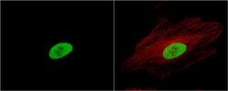
- Immunocytochemistry-Immunofluorescence analysis of Ku80 was performed in HeLa cells fixed in 4% paraformaldehyde at RT for 15 min. Green: Ku80 Polyclonal Antibody (Product # PA5-34758) diluted at 1:500. Red: phalloidin, a cytoskeleton marker.

- Submitted by
- Invitrogen Antibodies (provider)
- Main image

- Experimental details
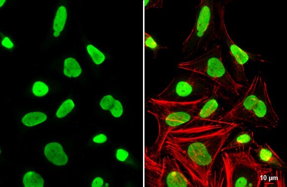
- Ku80 Polyclonal Antibody detects Ku80 protein at nucleus by immunofluorescent analysis. Sample: HeLa cells were fixed in 4% paraformaldehyde at RT for 15 min. Green: Ku80 stained by Ku80 Polyclonal Antibody (Product # PA5-34758) diluted at 1:1,000. Red: phalloidin, a cytoskeleton marker, diluted at 1:200. Scale bar= 10 µm.
- Submitted by
- Invitrogen Antibodies (provider)
- Main image
- Experimental details
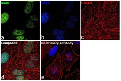
- Immunofluorescence analysis of Ku80 was performed using 70% confluent log phase HeLa cells. The cells were fixed with 4% paraformaldehyde for 10 minutes, permeabilized with 0.1% Triton™ X-100 for 15 minutes, and blocked with 2% BSA for 1 hour at room temperature. The cells were labeled with Ku80 Polyclonal Antibody (Product # PA5-34758) at 1:200 dilution in 0.1% BSA, incubated at 4 degree Celsius overnight and then with Goat anti-Rabbit IgG (H+L), Superclonal™ Recombinant Secondary Antibody, Alexa Fluor 488 conjugate (Product # A27034) at a dilution of 1:2000 for 45 minutes at room temperature (Panel a: green). Nuclei (Panel b: blue) were stained with SlowFade® Gold Antifade Mountant with DAPI (Product # S36938). F-actin (Panel c: red) was stained with Rhodamine Phalloidin (Product # R415, 1:300). Panel d represents the merged image showing staining in nucleus. Panel e represents control cells with no primary antibody to assess background. The images were captured at 60X magnification.

Supportive validation
- Submitted by
- Invitrogen Antibodies (provider)
- Main image
- Experimental details
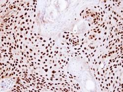
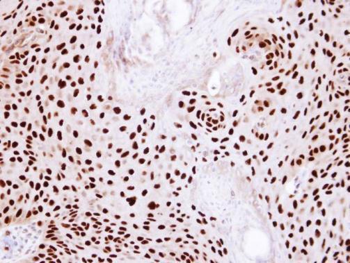
- Immunohistochemical analysis of paraffin-embedded Cal27 Xenograft, using Ku80 (XRCC5) (Product # PA5-34758) antibody at 1:100 dilution. Antigen Retrieval: EDTA based buffer, pH 8.0, 15 min.
Supportive validation
- Submitted by
- Invitrogen Antibodies (provider)
- Main image

- Experimental details
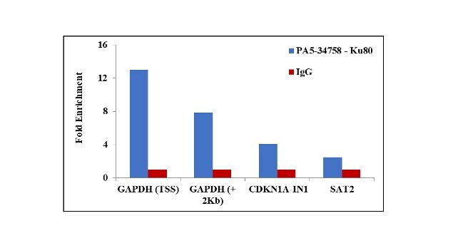
- Chromatin Immunoprecipitation (ChIP) assay of endogenous Ku80 protein using Anti-Ku80 Antibody: ChIP was performed using Anti-Ku80 Polyclonal Antibody (Product # PA5-34758, 2.5 µg) on sheared chromatin from camptothecin treated HeLa cells using the MAGnify ChIP System kit (Product # 49-2024). Normal Rabbit IgG was used as a negative IP control. The purified DNA was analyzed by qPCR using primers binding to GAPDH transcriptional start site, GAPDH gene body (+2Kb), CDKN1A intron 1 and SAT2 satellite repeats. Data is presented as fold enrichment of the antibody signal versus the negative control IgG using the comparative CT method.
Supportive validation
- Submitted by
- Invitrogen Antibodies (provider)
- Main image
- Experimental details
- Ku80 (XRCC5) antibody immunoprecipitates Ku80 protein in IP experiments. IP Sample: 1,000 µg HeLa whole cell lysate/extract A. 40 µg HeLa whole cell lysate/extract B. Control with 2.5 µg of preimmune rabbit IgG C. Immunoprecipitation of Ku80 protein by 2.5 µg of Ku80 antibody (Product # PA5-34758) 7.5% SDS-PAGE The immunoprecipitated Ku80 protein was detected by Ku80 antibody (Product # PA5-34758) diluted at 1:1,000.

- Submitted by
- Invitrogen Antibodies (provider)
- Main image

- Experimental details
- Ku80 (XRCC5) antibody immunoprecipitates Ku80 protein in IP experiments. IP Sample: 1,000 µg HeLa whole cell lysate/extract A. 40 µg HeLa whole cell lysate/extract B. Control with 2.5 µg of preimmune rabbit IgG C. Immunoprecipitation of Ku80 protein by 2.5 µg of Ku80 antibody (Product # PA5-34758) 7.5% SDS-PAGE The immunoprecipitated Ku80 protein was detected by Ku80 antibody (Product # PA5-34758) diluted at 1:1,000.