 Explore
 Validate
 Learn
Learn Western blot
 Immunohistochemistry
Antibody data
- Antibody Data
- Antigen structure
- References [1]
- Comments [0]
- Validations
- Western blot [1]
- Other assay [1]
Submit
Validation data
Reference
Comment
Report error
- Product number
- PA5-17819 - Provider product page

- Provider
- Invitrogen Antibodies
- Product name
- Phospho-PP1 alpha (Thr320) Polyclonal Antibody
- Antibody type
- Polyclonal
- Antigen
- Synthetic peptide
- Description
- It is not recommended to aliquot this antibody.
- Reactivity
- Human, Mouse, Rat
- Host
- Rabbit
- Isotype
- IgG
- Vial size
- 100 µL
- Concentration
- 165 µg/mL
- Storage
- -20°C
Submitted references The PP1 regulator PPP1R2 coordinately regulates AURKA and PP1 to control centrosome phosphorylation and maintain central spindle architecture.
Bresch AM, Yerich N, Wang R, Sperry AO
BMC molecular and cell biology 2020 Nov 25;21(1):84
BMC molecular and cell biology 2020 Nov 25;21(1):84
No comments: Submit comment
Supportive validation
- Submitted by
- Invitrogen Antibodies (provider)
- Main image

- Experimental details
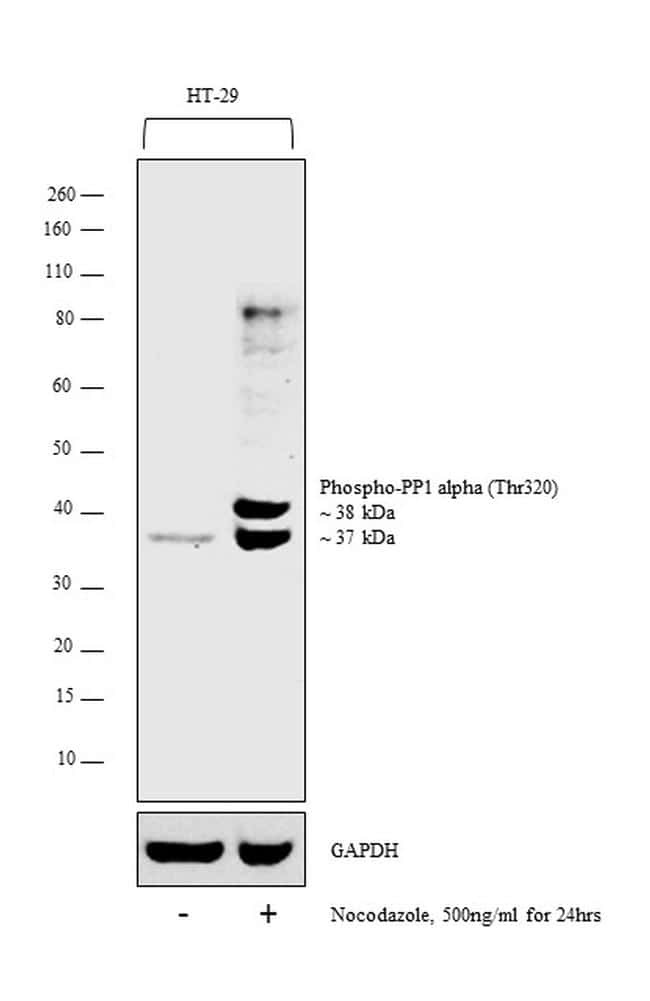
- Western blot analysis was performed on nuclear enriched cell extract (30 µg lysate) of HT-29 (Lane 1) and HT-29 treated with Nocodazole (500 ng/mL for 24 hours) (Lane 2). The blot was probed with Anti-Phospho-PP1 alpha (Thr320) Polyclonal Antibody (Product # PA5-17819, 1:1000 dilution) and detected by chemiluminescence using Goat anti-Rabbit IgG (H+L) Superclonal™ Secondary Antibody, HRP conjugate (Product # A27036, 0.25 µg/mL, 1:4000 dilution). A 37 and 38 kDa band corresponding to Phospho-PP1 alpha was detected in HT-29 upon Nocodazole treatment.
Supportive validation
- Submitted by
- Invitrogen Antibodies (provider)
- Main image
- Experimental details
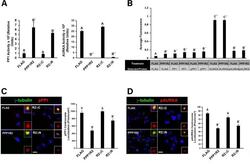
- Fig. 8 The effect of PPP1R2 on PP1 and AURKA activity at the centrosome was dependent on its C-terminus. a A graphical representation of the PP1 and AURKA activity of cells expressing each construct is shown. b Protein expression and phosphorylation levels of proteins indicated by the table below the graph were measured using ELISA. Cells were transfected with the indicated constructs, fixed, and labeled for gamma-tubulin (red) and either ( c ) phosphorylated PP1 (pPP1, green) or ( d ) phosphorylated AURKA (pAURKA, green) antibodies. pPP1 and pAURKA levels at the centrosome were quantified using Metamorph software and are displayed as graphs in C and D. Statistically significant differences ( p
