Explore
 Validate
 Learn
Learn Western blot
Antibody data
- Antibody Data
- Antigen structure
- References [0]
- Comments [0]
- Validations
- Western blot [4]
- Immunohistochemistry [1]
Submit
Validation data
Reference
Comment
Report error
- Product number
- PA5-40700 - Provider product page

- Provider
- Invitrogen Antibodies
- Product name
- SP3 Polyclonal Antibody
- Antibody type
- Polyclonal
- Antigen
- Synthetic peptide
- Description
- Peptide sequence: TAGINADGHL INTGQAMDSS DNSERTGERV SPDINETNTD TDLFVPTSSS
- Concentration
- 0.5 mg/mL
No comments: Submit comment
Supportive validation
- Submitted by
- Invitrogen Antibodies (provider)
- Main image

- Experimental details
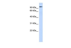
- Western blot analysis of human U2OS cells using an anti-SP3 polyclonal antibody (Product # PA5-40700).; Secondary Antibody: anti-Rabbit HRP.
- Submitted by
- Invitrogen Antibodies (provider)
- Main image
- Experimental details
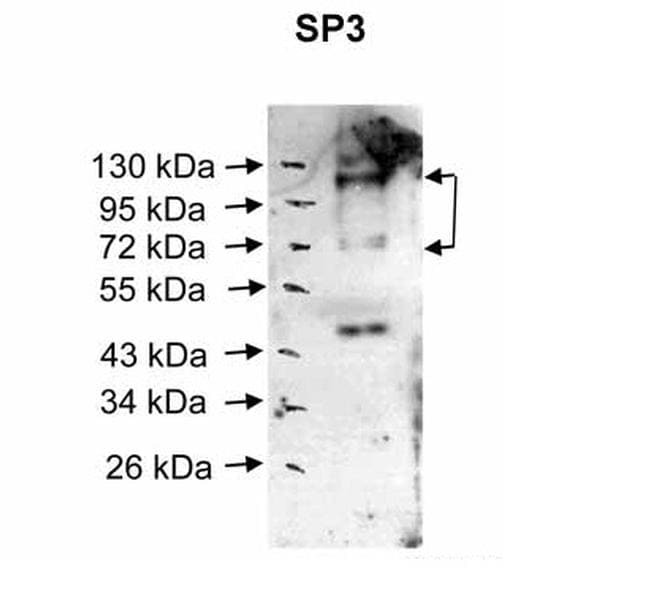
- Western blot analysis of human muscle cells using an anti-SP3 polyclonal antibody (Product # PA5-40700).

- Submitted by
- Invitrogen Antibodies (provider)
- Main image
- Experimental details
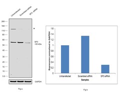
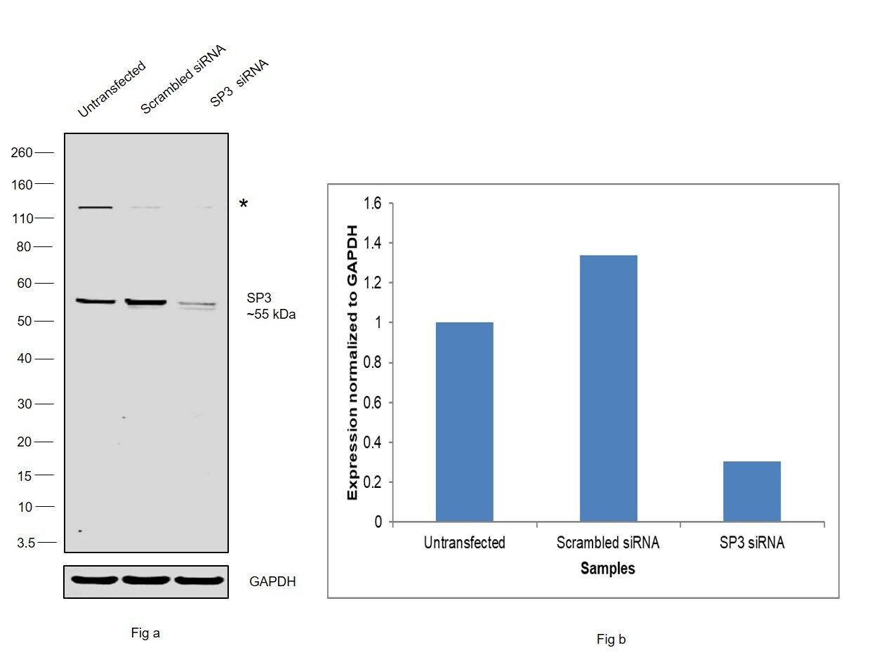
- Knockdown of SP3 was achieved by transfecting HeLa with SP3 specific siRNAs (Silencer® select Product # S13324, S13325). Western Blot analysis (Fig. a) was performed using whole cell extracts from the SP3 knockdown cells (lane 3), non-specific scrambled siRNA transfected cells (lane 2) and untransfected cells (lane 1). The Blot was probed with SP3 Polyclonal Antibody (Product # PA5-40700, 1:1000 dilution) and Goat anti-Rabbit IgG (H+L) Superclonal™ Secondary Antibody, HRP (Product # A27036, 0.25 µg/mL, 1:4000 dilution). Densitometric analysis of this western Blot is shown in histogram (Fig. b). Decrease in signal upon siRNA mediated knock down confirms that antibody is specific to SP3. Additional uncharacterized band was observed at ~120 kDa.
- Submitted by
- Invitrogen Antibodies (provider)
- Main image
- Experimental details
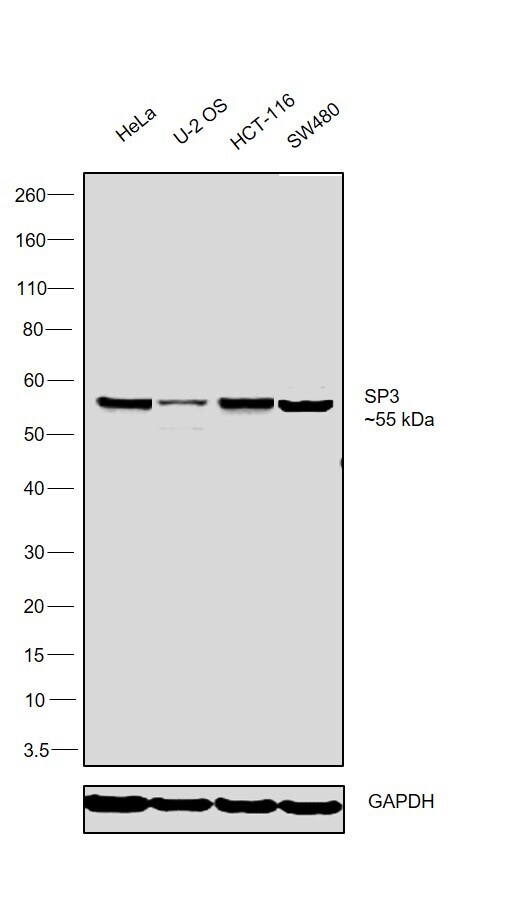
- Western Blot was performed using SP3 Polyclonal Antibody (Product # PA5-40700) and a ~55 kDa band corresponding to SP3 was observed across cell lines tested. Nuclear extracts (30 µg lysate) of HeLa (Lane 1), U-2 OS (Lane 2), HCT-116 (Lane 3) and SW480 (Lane 4) were electrophoresed using NuPAGE™ 4-12% Bis-Tris Protein Gel (Product # NP0321BOX). Resolved proteins were then transferred onto a Nitrocellulose membrane (Product # LC2001) by iBlot® 2 Dry Blotting System (Product # IB21001). The Blot was probed with the primary antibody (1:1000 dilution) and detected by chemiluminescence with Goat anti-Rabbit IgG (H+L) Superclonal™ Recombinant Secondary Antibody, HRP (Product # A27036, 1:4000 dilution) using the iBright FL 1000 (Product # A32752). Chemiluminescent detection was performed using Novex® ECL Chemiluminescent Substrate Reagent Kit (Product # WP20005).
Supportive validation
- Submitted by
- Invitrogen Antibodies (provider)
- Main image

- Experimental details
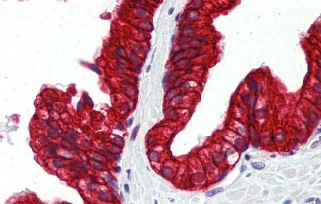
- Immunohistochemistry analysis of human prostate tissue using an anti-SP3 polyclonal antibody (Product # PA5-40700).