Explore
 Validate
 Learn
Learn Western blot
Antibody data
- Antibody Data
- Antigen structure
- References [0]
- Comments [0]
- Validations
- Western blot [4]
- Immunocytochemistry [3]
- Immunohistochemistry [1]
Submit
Validation data
Reference
Comment
Report error
- Product number
- PA5-54984 - Provider product page

- Provider
- Invitrogen Antibodies
- Product name
- TOP2B Polyclonal Antibody
- Antibody type
- Polyclonal
- Antigen
- Recombinant full-length protein
- Description
- Immunogen sequence: LWKEDLAAFV EELDKVESQE REDVLAGMSG KAIKGKVGKP KVKKLQLEET MPSPYGRRII PEITAMKADA SKKLLKKKKG DLDTAAVKVE FDEEFSGAPV EGAGEEALTP SVPINKGPKP KREKKEPGTR VRKTPTSSGK PSA
- Concentration
- 0.2 mg/mL
No comments: Submit comment
Supportive validation
- Submitted by
- Invitrogen Antibodies (provider)
- Main image
- Experimental details
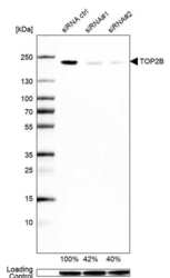
- Western blot analysis of TOP2B in U2OS cells transfected with control siRNA, target specific siRNA probe #1 and #2, using a TOP2B Polyclonal Antibody (Product # PA5-54984). Remaining relative intensity is presented. Loading control: Anti-GAPDH.

- Submitted by
- Invitrogen Antibodies (provider)
- Main image

- Experimental details
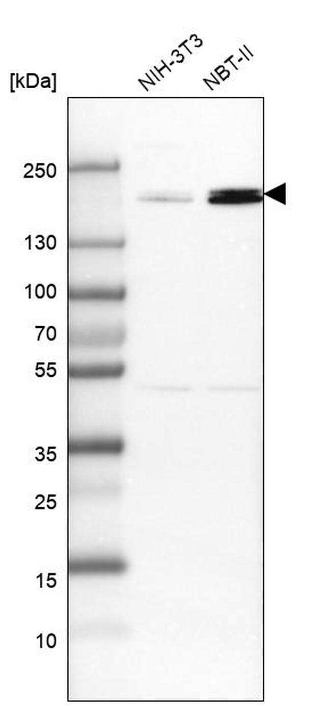
- Western blot analysis of TOP2B in mouse cell line NIH-3T3 and rat cell line NBT-II using a TOP2B Polyclonal Antibody (Product # PA5-54984).
- Submitted by
- Invitrogen Antibodies (provider)
- Main image

- Experimental details
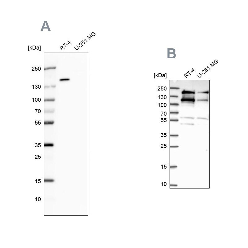
- Western blot analysis of TOP2B using TOP2B Polyclonal Antibody (Product # PA5-54984) (A) shows similar pattern to an independent TOP2B Polyclonal Antibody (B).
- Submitted by
- Invitrogen Antibodies (provider)
- Main image

- Experimental details
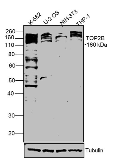
- Western blot was performed using TOP2B Polyclonal Antibody (Product # PA5-54984) and a 160 kDa band corresponding to TOP2B was observed in all tested cell lines along with few uncharacterized bands. Whole cell extracts (30 µg lysate) of K-562 (Lane 1), U-2 OS (Lane 2), NIH-3T3 (Lane 3), THP-1 (Lane 4) were electrophoresed using NuPAGE® 4-12 % Bis-Tris gel (Product # NP0321BOX). Resolved proteins were then transferred onto a nitrocellulose membrane (Product # IB23001) by iBlot® 2 Dry Blotting System (Product # IB21001). The blot was probed with the primary antibody (1:1000 dilution) and detected by chemiluminescence with Goat anti-Rabbit IgG (H+L) Superclonal™ Recombinant Secondary Antibody, HRP (Product # A27036, 1:4000 dilution) using the iBright FL 1000 (Product # A32752). Chemiluminescent detection was performed using Novex® ECL Chemiluminescent Substrate Reagent Kit (Product # WP20005).
Supportive validation
- Submitted by
- Invitrogen Antibodies (provider)
- Main image
- Experimental details
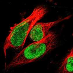
- Immunofluorescent staining of TOP2B in human cell line U-251 MG shows positivity in nucleus but excluded from the nucleoli. Samples were probed using a TOP2B Polyclonal Antibody (Product # PA5-54984).

- Submitted by
- Invitrogen Antibodies (provider)
- Main image
- Experimental details
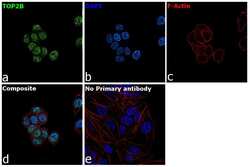
- Immunofluorescence analysis of TOP2B was performed using U-2 OS cells. The cells were fixed with 4% paraformaldehyde for 10 minutes, permeabilized with 0.1% Triton™ X-100 for 15 minutes, and blocked with 2% BSA for 1 hour at room temperature. The cells were labeled with TOP2B Polyclonal Antibody (Product # PA5-54984) at 2.5 µg/mL concentration in 0.1% BSA and incubated overnight at 4 degree and then labeled with Goat anti-Rabbit IgG (H+L) Superclonal™ Recombinant Secondary Antibody, Alexa Fluor® 488 (Product # A27034, 1:2000 dilution) for 45 minutes at room temperature (Panel a: green) in U-2 OS cells. Nuclei (Panel b: blue) were stained with ProLong™ Diamond Antifade Mountant with DAPI (Product # P36962). F-actin (Panel c: red) was stained with Rhodamine Phalloidin (Product # R415, 1:300). Panel d represents the merged image of U-2 OS cells. Panel f represents control cells with no primary antibody to assess background. The images were captured at 60X magnification.

- Submitted by
- Invitrogen Antibodies (provider)
- Main image

- Experimental details
- Immunofluorescence analysis of TOP2B was performed using U-2 OS cells. The cells were fixed with 4% paraformaldehyde for 10 minutes, permeabilized with 0.1% Triton™ X-100 for 15 minutes, and blocked with 2% BSA for 1 hour at room temperature. The cells were labeled with TOP2B Polyclonal Antibody (Product # PA5-54984) at 2.5 µg/mL concentration in 0.1% BSA and incubated overnight at 4 degree and then labeled with Goat anti-Rabbit IgG (H+L) Superclonal™ Recombinant Secondary Antibody, Alexa Fluor® 488 (Product # A27034, 1:2000 dilution) for 45 minutes at room temperature (Panel a: green) in U-2 OS cells. Nuclei (Panel b: blue) were stained with ProLong™ Diamond Antifade Mountant with DAPI (Product # P36962). F-actin (Panel c: red) was stained with Rhodamine Phalloidin (Product # R415, 1:300). Panel d represents the merged image of U-2 OS cells. Panel f represents control cells with no primary antibody to assess background. The images were captured at 60X magnification.

Supportive validation
- Submitted by
- Invitrogen Antibodies (provider)
- Main image

- Experimental details
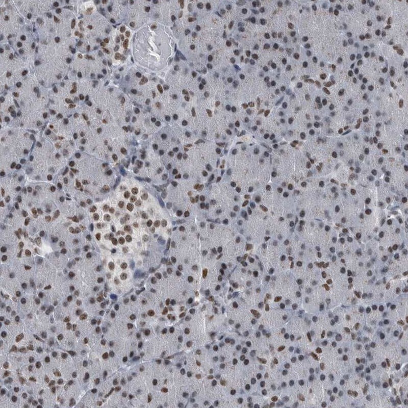
- Immunohistochemical staining of TOP2B in human pancreas using a TOP2B Polyclonal Antibody (Product # PA5-54984) shows moderate nuclear positivity.