Explore
 Validate
 Learn
Learn Western blot
Antibody data
- Antibody Data
- Antigen structure
- References [6]
- Comments [0]
- Validations
- Western blot [4]
- Immunocytochemistry [4]
- Flow cytometry [1]
- Other assay [4]
Submit
Validation data
Reference
Comment
Report error
- Product number
- MA1-19421 - Provider product page

- Provider
- Invitrogen Antibodies
- Product name
- gamma Tubulin Monoclonal Antibody (TU-30)
- Antibody type
- Monoclonal
- Antigen
- Synthetic peptide
- Description
- This antibody recognizes the C-terminal peptide sequence EYHAATRPDYISWGTQ (aa 434-449) of gamma-tubulin.
- Antibody clone number
- TU-30
- Concentration
- 1 mg/mL
Submitted references Pathogenic LRRK2 regulates ciliation probability upstream of tau tubulin kinase 2 via Rab10 and RILPL1 proteins.
WDR62 regulates spindle dynamics as an adaptor protein between TPX2/Aurora A and katanin.
Loss of AMPKalpha1 Triggers Centrosome Amplification via PLK4 Upregulation in Mouse Embryonic Fibroblasts.
Drp1 Controls Effective T Cell Immune-Surveillance by Regulating T Cell Migration, Proliferation, and cMyc-Dependent Metabolic Reprogramming.
Plk1-dependent recruitment of gamma-tubulin complexes to mitotic centrosomes involves multiple PCM components.
Nuclear gamma-tubulin during acentriolar plant mitosis.
Sobu Y, Wawro PS, Dhekne HS, Yeshaw WM, Pfeffer SR
Proceedings of the National Academy of Sciences of the United States of America 2021 Mar 9;118(10)
Proceedings of the National Academy of Sciences of the United States of America 2021 Mar 9;118(10)
WDR62 regulates spindle dynamics as an adaptor protein between TPX2/Aurora A and katanin.
Huang J, Liang Z, Guan C, Hua S, Jiang K
The Journal of cell biology 2021 Aug 2;220(8)
The Journal of cell biology 2021 Aug 2;220(8)
Loss of AMPKalpha1 Triggers Centrosome Amplification via PLK4 Upregulation in Mouse Embryonic Fibroblasts.
Zhao Q, Coughlan KA, Zou MH, Song P
International journal of molecular sciences 2020 Apr 16;21(8)
International journal of molecular sciences 2020 Apr 16;21(8)
Drp1 Controls Effective T Cell Immune-Surveillance by Regulating T Cell Migration, Proliferation, and cMyc-Dependent Metabolic Reprogramming.
Simula L, Pacella I, Colamatteo A, Procaccini C, Cancila V, Bordi M, Tregnago C, Corrado M, Pigazzi M, Barnaba V, Tripodo C, Matarese G, Piconese S, Campello S
Cell reports 2018 Dec 11;25(11):3059-3073.e10
Cell reports 2018 Dec 11;25(11):3059-3073.e10
Plk1-dependent recruitment of gamma-tubulin complexes to mitotic centrosomes involves multiple PCM components.
Haren L, Stearns T, Lüders J
PloS one 2009 Jun 19;4(6):e5976
PloS one 2009 Jun 19;4(6):e5976
Nuclear gamma-tubulin during acentriolar plant mitosis.
Binarová P, Cenklová V, Hause B, Kubátová E, Lysák M, Dolezel J, Bögre L, Dráber P
The Plant cell 2000 Mar;12(3):433-42
The Plant cell 2000 Mar;12(3):433-42
No comments: Submit comment
Supportive validation
- Submitted by
- Invitrogen Antibodies (provider)
- Main image
- Experimental details
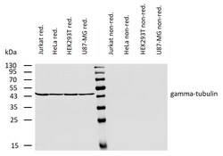
- Western blotting analysis of human gamma-tubulin using mouse monoclonal antibody TU-30 on lysates of various cell lines under reducing and non-reducing conditions. Nitrocellulose membrane was probed with 2µg/mL of mouse anti-gamma-tubulin monoclonal antibody (Product # MA1-19421) followed by IRDye800-conjugated anti-mouse secondary antibody. A specific band was detected for gamma-tubulin at approximately 46kDa.
- Submitted by
- Invitrogen Antibodies (provider)
- Main image

- Experimental details
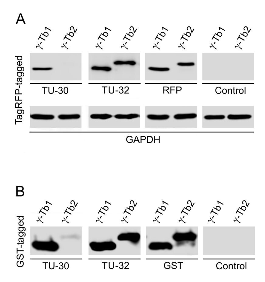
- Western blotting analysis of differential reactivity of monoclonal antibodies to γ-tubulin with human γ-tubulin isotypes. (A) Immunoblots of total cell lysates from SH-SY5Y cells, expressing TagRFP-tagged human γ-tubulin 1 (γ-Tb1) or γ-tubulin 2 (γ-Tb2), probed with antibodies to γ-tubulin (TU-30) (Product # MA1-19421), γ-tubulin (TU-32), TagRFP (RFP) and GAPDH. In control samples, only secondary anti-mouse antibody was applied. (B) Immunoblots of immobilized GST-tagged human C-terminal regions (a.a. 362-451) of γ-Tb1 or γ-Tb2 probed with antibodies to γ-tubulin (TU-30, TU-32) and GST. In control samples, only secondary anti-mouse antibody was applied.
- Submitted by
- Invitrogen Antibodies (provider)
- Main image
- Experimental details
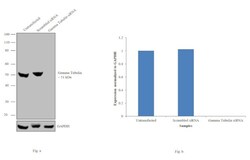
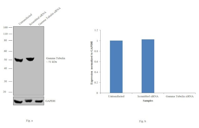
- Knockdown of gamma Tubulin was achieved by transfecting HeLa cells with gamma Tubulin specific siRNAs (Silencer® select Product # s14502). Western blot analysis (Fig. a) was performed using membrane extracts from the gamma Tubulin knockdown cells (lane 3), non-specific scrambled siRNA transfected cells (lane 2) and untransfected cells (lane 1). The blots were probed with gamma Tubulin Monoclonal Antibody (TU30) (Product # MA1-19421, 1:1000 dilution) and Goat anti-Mouse IgG (H+L) Superclonal™ Secondary Antibody, HRP conjugate (Product # A28177, 0.25 µg/mL, 1:4000 dilution). Densitometric analysis of this western blot is shown in histogram (Fig. b). Decrease in signal upon siRNA mediated knock down confirms that antibody is specific to gamma Tubulin.
- Submitted by
- Invitrogen Antibodies (provider)
- Main image
- Experimental details
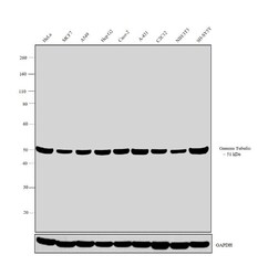
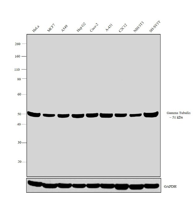
- Western blot analysis was performed on whole cell extracts (30 µg lysate) of HeLa (Lane 1), MCF7 (Lane 2), A549 (Lane 3), Hep G2 (Lane 4), Caco-2 (Lane 5), A-431 (Lane 6), C2C12 (Lane 7), NIH/3T3 (Lane 8) and SH-SY5Y (Lane 9). The blot was probed with Anti-gamma Tubulin Monoclonal Antibody (TU30) (Product # MA1-19421, 1:1000 dilution) and detected by chemiluminescence using Goat anti Mouse IgG (H+L) Superclonal™ Secondary Antibody, HRP conjugate (Product # A28177, 0.25 µg/mL, 1:4000 dilution). A 51 kDa band corresponding to gamma Tubulin was observed across all cell lines tested.
Supportive validation
- Submitted by
- Invitrogen Antibodies (provider)
- Main image

- Experimental details
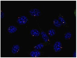
- Immunofluorescent analysis of gamma Tubulin using a monoclonal antibody (Product # MA1-19421).
- Submitted by
- Invitrogen Antibodies (provider)
- Main image

- Experimental details
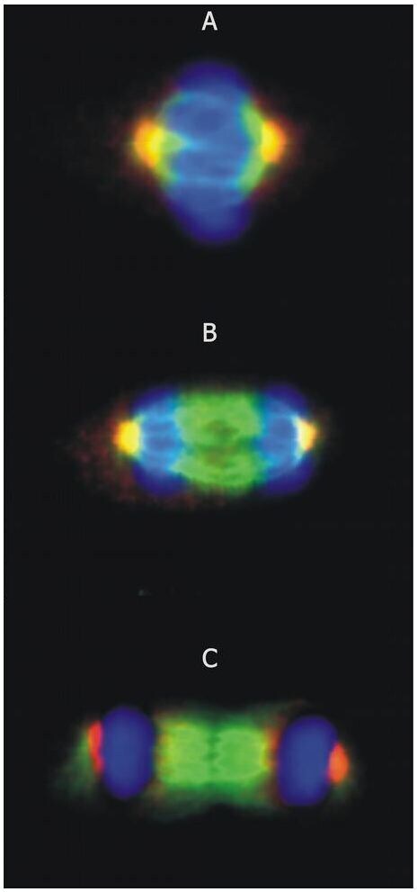
- Immunocytochemistry staining of microtubular networks in 3T3 mouse fibroblasts. A – metaphase; B – anaphase; C - telophase. Gamma-tubulin (red) stained with anti-gamma-tubulin (TU-30) Monoclonal antibody (Product # MA1-19421), alpha-tubulin (green) with polyclonal anti-alpha-tubulin antibody and nuclei with DAPI (blue).
- Submitted by
- Invitrogen Antibodies (provider)
- Main image
- Experimental details
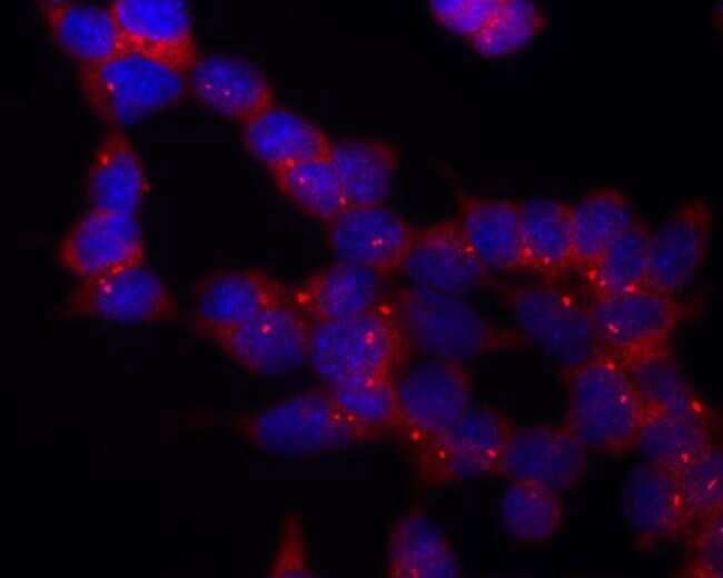
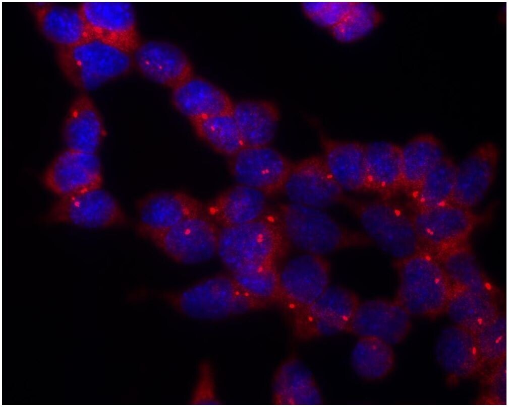
- Immunocytochemistry staining of murine fibroblasts using anti-gamma-tubulin (TU-30; direct conjugate with Dyomics 547, red) Monoclonal antibody (Product # MA1-19421). Nuclei were stained with DAPI (blue).

- Submitted by
- Invitrogen Antibodies (provider)
- Main image

- Experimental details
- Immunocytochemistry staining of P19X1 mouse embryonal carcinoma cell line using anti-gamma-tubulin (TU-30) Monoclonal antibody (Product # MA1-19421), detection by secondary antibody Goat anti-mouse Cy3. Nuclei were stained with DAPI (blue).
Supportive validation
- Submitted by
- Invitrogen Antibodies (provider)
- Main image
- Experimental details
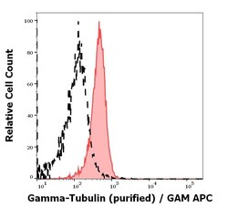
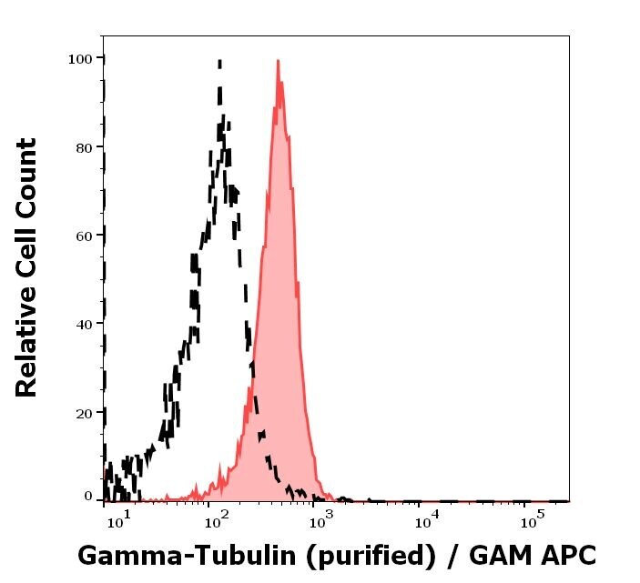
- Separation of MCF-7 cells stained using anti-gamma-Tubulin (TU-30) purified Monoclonal antibody (Product # MA1-19421) (concentration in sample 9 µg/mL, GAM APC, red-filled) from MCF-7 cells unstained by primary antibody (GAM APC, black-dashed) in flow cytometry analysis (intracellular staining).
Supportive validation
- Submitted by
- Invitrogen Antibodies (provider)
- Main image

- Experimental details
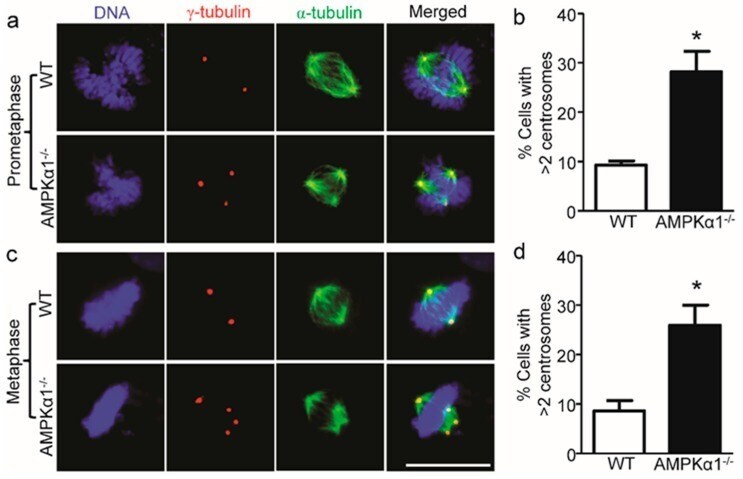
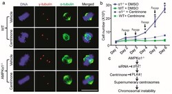
- Figure 2 AMPKalpha1 deletion confers centrosome amplification in MEFs. ( a ) Representative images of centrosome morphologies of WT and AMPKalpha1 -/- MEFs (scale bar = 20 mum) in the prometaphase of the cell cycle are shown. Centrosomes, spindles, and DNA were co-stained with anti-gamma-tubulin antibody (red), anti-alpha-tubulin antibody (green), and 4.6-diamidino-2-phenylindole (DAPI, blue), respectively, and visualized by fluorescence microscope; ( b ) Quantification data for the percentage of MEFs containing >2 centrosomes are presented. n = 5, * p < 0.01, AMPKalpha1 -/- vs. WT; ( c ) Representative images of centrosome morphologies of WT and AMPKalpha1 -/- MEFs (scale bar = 20 mum) in the metaphase of the cell cycle are shown; ( d ) Percentage of cells with >2 centrosomes. n = 5, * p < 0.01, AMPKalpha1 -/- vs. WT.
- Submitted by
- Invitrogen Antibodies (provider)
- Main image

- Experimental details
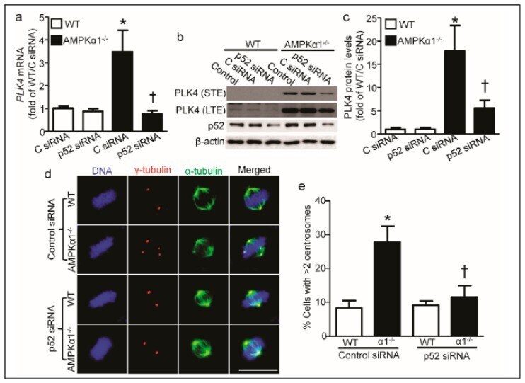
- Figure 4 PLK4 elevation is p52-mediated. ( a ) p52 knockdown blocked the PLK4 mRNA upregulation in AMPKalpha1 -/- MEFs. n = 3, * p < 0.01, vs. C siRNA/WT; + p < 0.01, vs. C siRNA/AMPKalpha1 -/- ; ( b ) Representative Western blotting showed p52 siRNA decreased the PLK4 protein elevated in AMPKalpha1 -/- MEFs. STE: short-term exposure; LTE: long-term exposure; ( c ) Quantification data of PLK4 protein levels for (b); n = 4, * p < 0.01, vs. C siRNA/WT; + p < 0.01, vs. C siRNA/AMPKalpha1 -/- ; ( d ) MEFs were treated with p52 siRNA or control siRNA for 72 h before the start of the experiment. Cells were treated with 20 ng/mL nocodazole for 4 hours to depolymerize microtubules. Centrosomes, spindles, and DNA were co-stained with anti-gamma-tubulin antibody (red), anti-alpha-tubulin antibody (green), and 4.6-diamidino-2-phenylindole (DAPI, blue), respectively, and visualized by fluorescence microscope (scale bar = 20 mum). Representative images are shown; ( e ) Quantification data represent the mean +- S.D. from three separate experiments. * p < 0.01, vs. WT/control siRNA; + p < 0.01, vs. AMPKalpha1 -/- /control siRNA. C siRNA: control siRNA.
- Submitted by
- Invitrogen Antibodies (provider)
- Main image
- Experimental details
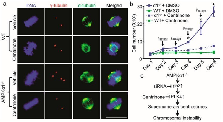
- Figure 5 PLK4 inhibition reverses centrosome amplification and cell hyperproliferation in AMPKalpha1-deleted MEFs. ( a ) PLK4 inhibition by Centrinone blocked centrosome amplification in AMPKalpha1 -/- MEFs. MEFs were treated with Centrinone (125 nM) or vehicle (DMSO) for 16 h. The cells were treated with nocodazole (20 ng/mL) for 4 hours to depolymerize the microtubules. Then, centrosomes, spindles, and DNA were co-stained with anti-gamma-tubulin antibody (red), anti-alpha-tubulin antibody (green), and 4.6-diamidino-2-phenylindole (DAPI, blue), respectively, and visualized by fluorescence microscope (scale bar = 20 mum); ( b ) PLK4 inhibition restrained cellular hyperproliferation in AMPKalpha1 -/- MEFs. MEF cells were seeded into 10-cm tissue culture dishes at 1 x 10 5 cells/dish. Centrinone was added at 125 nM. Every day, cells were harvested, counted using a Bio-Rad cell counter, and re-seeded into new dishes. Quantification data represent the mean +- S.D. from three separate experiments. * p < 0.01, vs. alpha1 -/- + Centrinone; ( c ) A scheme illustrating the role of p52-PLK4 in AMPKalpha1 deletion-mediated centrosome amplification. P52 transcriptionally regulates PLK4, which contributes to centrosome amplification and the consequent chromosome instability.
- Submitted by
- Invitrogen Antibodies (provider)
- Main image
- Experimental details
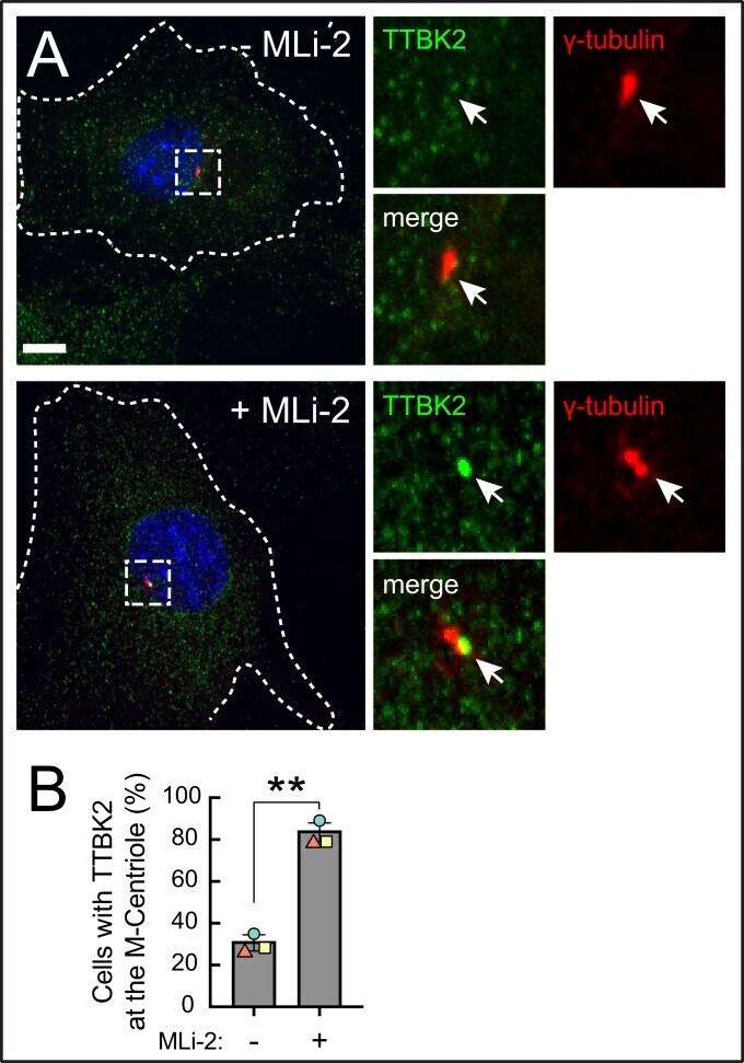
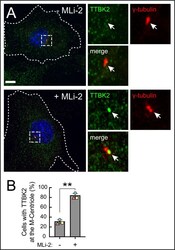
- Fig. 5. Pathogenic LRRK2 activity blocks TTBK2 recruitment to the mother centriole. ( A ) R1441C LRRK2 MEF cells stably expressing GFP-TTBK2 were starved for 24 h +- MLi-2. Cells were then fixed with cold methanol and stained with rabbit anti-TTBK2 (green) and mouse anti-gamma-tubulin (red) antibodies. At right are enlarged regions boxed in the larger images at left. ( B ) Percent of cells with TTBK2 at the centrosome, marked by gamma-tubulin staining. Values represent the mean and SEM of three independent experiments ( n > 60 in each experiment). Significance was determined by the paired t test; * P = 0.004. (Scale bars, 10 um.)