Explore
 Validate
 Learn
Learn Western blot
Antibody data
- Antibody Data
- Antigen structure
- References [1]
- Comments [0]
- Validations
- Western blot [4]
- Immunocytochemistry [1]
- Immunoprecipitation [1]
- Immunohistochemistry [2]
Submit
Validation data
Reference
Comment
Report error
- Product number
- GTX113124 - Provider product page

- Provider
- GeneTex
- Proper citation
- GeneTex Cat#GTX113124, RRID:AB_11165236
- Product name
- AHR antibody
- Antibody type
- Polyclonal
- Reactivity
- Human, Mouse, Rat
- Host
- Rabbit
- Storage
- Keep as concentrated solution. Aliquot and store at -20°C or below. Avoid multiple freeze-thaw cycles.
Submitted references Resolvin D1 down-regulates CYP1A1 and PTGS2 gene in the HUVEC cells treated with benzo(a)pyrene.
Gdula-Argasińska J, Czepiel J, Totoń-Żurańska J, Jurczyszyn A, Wołkow P, Librowski T, Perucki W
Pharmacological reports : PR 2016 Oct;68(5):939-44
Pharmacological reports : PR 2016 Oct;68(5):939-44
No comments: Submit comment
Supportive validation
- Submitted by
- GeneTex (provider)
- Main image

- Experimental details
- Sample (30 ug of whole cell lysate) A: 293T B: A431 7.5% SDS PAGE GTX113124 diluted at 1:1000
- Validation comment
- WB

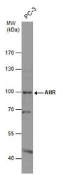
- Submitted by
- GeneTex (provider)
- Main image

- Experimental details
- AHR antibody detects AHR protein by western blot analysis.A. 30 ?g PC-3 whole cell lysate/extract7.5 % SDS-PAGEAHR antibody (GTX113124) dilution: 1:500
- Validation comment
- WB
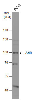
- Submitted by
- GeneTex (provider)
- Main image
- Experimental details
- Whole cell extract (30 μg) was separated by 7.5% SDS-PAGE, and the membrane was blotted with AHR antibody (GTX113124) diluted at 1:500.
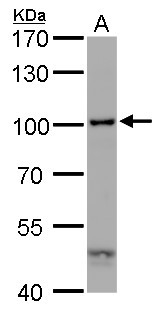
- Submitted by
- GeneTex (provider)
- Main image
- Experimental details
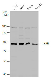
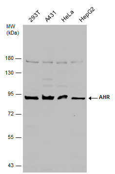
- Various whole cell extracts (30 μg) were separated by 7.5% SDS-PAGE, and the membrane was blotted with AHR antibody (GTX113124) diluted at 1:1000.
Supportive validation
- Submitted by
- GeneTex (provider)
- Main image

- Experimental details
- AHR antibody detects AHR protein at cytoplasm and nucleus by immunofluorescent analysis. Sample: HeLa cells were fixed in 4% paraformaldehyde at RT for 15 min.Green: AHR protein stained by AHR antibody (GTX113124) diluted at 1:500.Blue: Hoechst 33342 staining.

Supportive validation
- Submitted by
- GeneTex (provider)
- Main image
- Experimental details
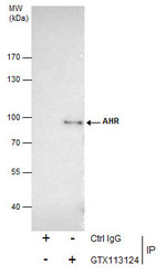
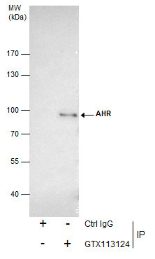
- Immunoprecipitation of AHR protein from 293T whole cell extracts using 5 £gg of AHR antibody (GTX113124).Western blot analysis was performed using AHR antibody (GTX113124).EasyBlot anti-Rabbit IgG (GTX221666-01) was used as a secondary reagent.
Supportive validation
- Submitted by
- GeneTex (provider)
- Main image
- Experimental details
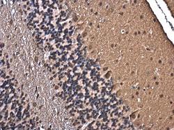
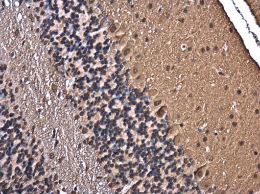
- AHR antibody detects AHR protein at nucleus in mouse brain by immunohistochemical analysis. Sample: Paraffin-embedded mouse brain. AHR antibody (GTX113124) diluted at 1:500.
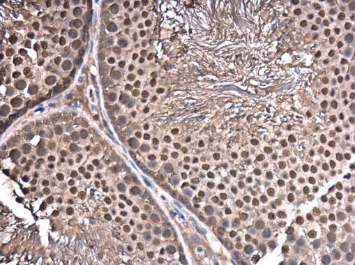
- Submitted by
- GeneTex (provider)
- Main image
- Experimental details
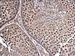
- AHR antibody detects AHR protein at nucleus in rat testis by immunohistochemical analysis. Sample: Paraffin-embedded rat testis. AHR antibody (GTX113124) diluted at 1:500.