Explore
 Validate
 Learn
Learn Western blot
Antibody data
- Antibody Data
- Antigen structure
- References [0]
- Comments [0]
- Validations
- Western blot [8]
- Immunocytochemistry [1]
- Immunohistochemistry [1]
Submit
Validation data
Reference
Comment
Report error
- Product number
- PA5-32194 - Provider product page

- Provider
- Invitrogen Antibodies
- Product name
- CTGF Polyclonal Antibody
- Antibody type
- Polyclonal
- Antigen
- Recombinant protein fragment
- Description
- Recommended positive controls: HUVEC, Rat bladder, CTGF-transfected 293T.
- Concentration
- 0.65 mg/mL
No comments: Submit comment
Supportive validation
- Submitted by
- Invitrogen Antibodies (provider)
- Main image
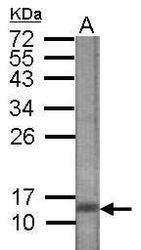
- Experimental details
- Western blot analysis of CTGF in 100 ng of 15 kDa recombinant CTGF using a CTGF polyclonal antibody (Product # PA5-32194) at a dilution of 1:90,000.
- Submitted by
- Invitrogen Antibodies (provider)
- Main image
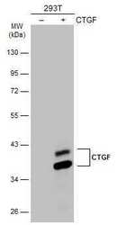
- Experimental details
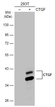
- Western Blot analysis of CTGF was performed by separating 30 µg of non-transfected (–) and transfected (+) 293T whole cell extracts by 10% SDS-PAGE. Proteins were transferred to a membrane and probed with a CTGF Polyclonal Antibody (Product # PA5-32194) at a dilution of 1:1000. The HRP-conjugated anti-rabbit IgG antibody was used to detect the primary antibody.

- Submitted by
- Invitrogen Antibodies (provider)
- Main image
- Experimental details
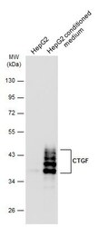
- Western blot analysis of CTGF was performed by separating 30 µg of HepG2 whole cell extract and conditioned medium by 10% SDS-PAGE. Proteins were transferred to a membrane and probed with a CTGF Polyclonal Antibody (Product # PA5-32194) at a dilution of 1:1000. The HRP-conjugated anti-rabbit IgG antibody was used to detect the primary antibody.

- Submitted by
- Invitrogen Antibodies (provider)
- Main image

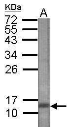
- Experimental details
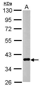
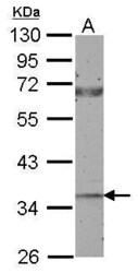
- Western Blot using CTGF Polyclonal Antibody (Product # PA5-32194). Sample (30 µg of whole cell lysate). Lane A: HUVEC. 10% SDS PAGE. CTGF Polyclonal Antibody (Product # PA5-32194) diluted at 1:1,000.
- Submitted by
- Invitrogen Antibodies (provider)
- Main image

- Experimental details
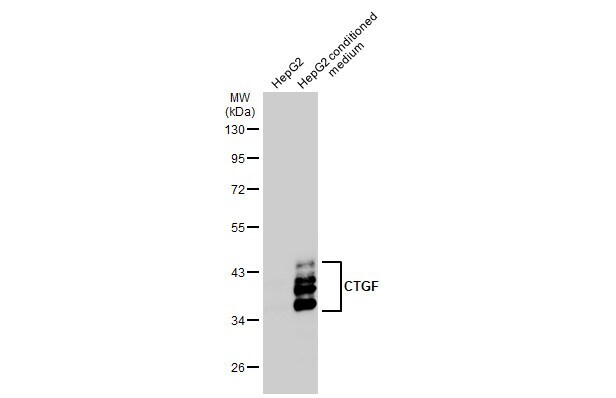
- Western Blot using CTGF Polyclonal Antibody (Product # PA5-32194). HepG2 conditioned medium (30 µg) were separated by 10% SDS-PAGE, and the membrane was blotted with CTGF Polyclonal Antibody (Product # PA5-32194) diluted at 1:1,000. The HRP-conjugated anti-rabbit IgG antibody was used to detect the primary antibody.
- Submitted by
- Invitrogen Antibodies (provider)
- Main image
- Experimental details
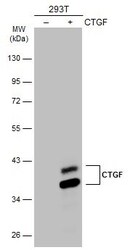
- Western Blot analysis of CTGF was performed by separating 30 µg of non-transfected (–) and transfected (+) 293T whole cell extracts by 10% SDS-PAGE. Proteins were transferred to a membrane and probed with a CTGF Polyclonal Antibody (Product # PA5-32194) at a dilution of 1:1000. The HRP-conjugated anti-rabbit IgG antibody was used to detect the primary antibody.
- Submitted by
- Invitrogen Antibodies (provider)
- Main image
- Experimental details
- Western Blot using CTGF Polyclonal Antibody (Product # PA5-32194). Sample (50 µg of whole cell lysate). Lane A: Rat bladder. 10% SDS PAGE. CTGF Polyclonal Antibody (Product # PA5-32194) diluted at 1:500.

- Submitted by
- Invitrogen Antibodies (provider)
- Main image
- Experimental details
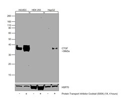
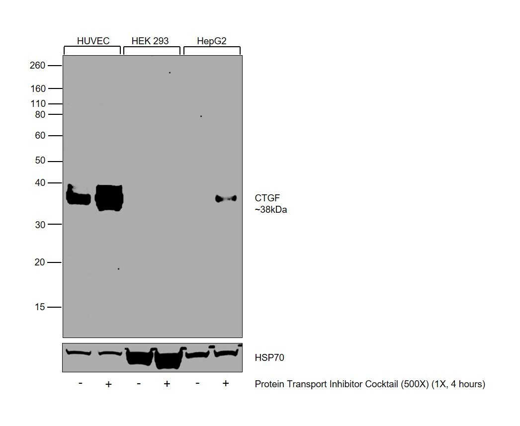
- Western Blot was performed using Anti-CTGF Polyclonal Antibody (Product # PA5-32194) and a 38 kDa band corresponding to Connective tissue growth factor was observed across all the tested cell lines. Whole cell extracts (30 µg lysate) of HUVEC (Lane 1), HUVEC (Protein Transport Inhibitor Cocktail (500X), 1X, 4 hours)(Lane 2), HEK-293 (Lane 3), HEK-293 (Protein Transport Inhibitor Cocktail (500X), 1X, 4 hours)(Lane 4), Hep G2 (Lane 5), Hep G2 (Protein Transport Inhibitor Cocktail (500X), 1X, 4 hours)(Lane 6) were electrophoresed using NuPAGE™ 10% Bis-Tris Protein Gel (Product # NP0302BOX). Resolved proteins were then transferred onto a Nitrocellulose membrane (Product # IB23001) by iBlot® 2 Dry Blotting System (Product # IB21001). The Blot was probed with the primary antibody (1:1000) and detected by chemiluminescence with Goat anti-Rabbit IgG (H+L) Superclonal™ Recombinant Secondary Antibody, HRP (Product # A27036, 1:4000) using the iBright FL 1000 (Product # A32752). Chemiluminescent detection was performed using Novex® ECL Chemiluminescent Substrate Reagent Kit (Product # WP20005).
Supportive validation
- Submitted by
- Invitrogen Antibodies (provider)
- Main image
- Experimental details
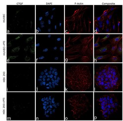
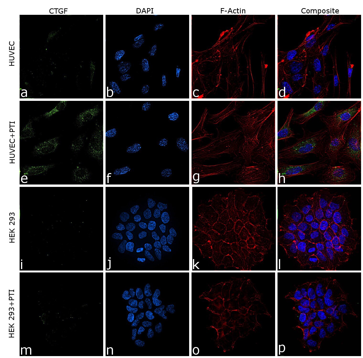
- Immunofluorescence analysis of CTGF was performed using 70% confluent log phase HUVEC cells. The cells were fixed with 4% paraformaldehyde for 10 minutes, permeabilized with 0.1% Triton™ X-100 for 15 minutes, and blocked with 2% BSA for 45 minutes at room temperature. The cells were labeled with CTGF Polyclonal Antibody (Product # PA5-32194) at 5 µg/mL in 0.1% BSA, incubated at 4 degree celsius overnight and then labeled with Donkey anti-Rabbit IgG (H+L) Highly Cross-Adsorbed Secondary Antibody, Alexa Fluor Plus 488 (Product # A32790), (1:2000), for 45 minutes at room temperature (Panel a: Green). Nuclei (Panel b: Blue) were stained with ProLong™ Diamond Antifade Mountant with DAPI (Product # P36962). F-actin (Panel c: Red) was stained with Rhodamine Phalloidin (Product # R415, 1:300). Panel d represents the merged image showing cytoplasmic localization. Panel (a-d) shows representative HUVEC control cell, whereas Panel (e-h) represent HUVEC with PTI treatment. Similarly, panel (i-l) represent HEK 293 control cells and Panel (m-p) represent HEK 293 with PTI treatment. The images were captured at 60X magnification.
Supportive validation
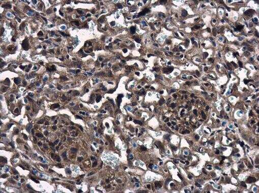
- Submitted by
- Invitrogen Antibodies (provider)
- Main image
- Experimental details
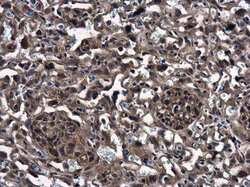
- Immunohistochemistry (Paraffin) analysis of CTGF was performed in paraffin-embedded mouse placenta tissue using CTGF Polyclonal Antibody (Product # PA5-32194) at a dilution of 1:500.