 Explore
 Validate
 Learn
Learn Western blot
Antibody data
- Antibody Data
- Antigen structure
- References [1]
- Comments [0]
- Validations
- Western blot [6]
- Immunocytochemistry [5]
- Immunohistochemistry [7]
Submit
Validation data
Reference
Comment
Report error
- Product number
- PA5-29705 - Provider product page

- Provider
- Invitrogen Antibodies
- Product name
- GRP78 Polyclonal Antibody
- Antibody type
- Polyclonal
- Antigen
- Recombinant protein fragment
- Description
- Recommended positive controls: 293T, A431, HeLa, HepG2, NIH-3T3, JC, BCL-1, PC-12, Rat2.
- Concentration
- 0.56 mg/mL
Submitted references An excess dietary vitamin E concentration does not influence Nrf2 signaling in the liver of rats fed either soybean oil or salmon oil.
Eder K, Siebers M, Most E, Scheibe S, Weissmann N, Gessner DK
Nutrition & metabolism 2017;14:71
Nutrition & metabolism 2017;14:71
No comments: Submit comment
Supportive validation
- Submitted by
- Invitrogen Antibodies (provider)
- Main image
- Experimental details
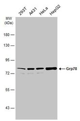
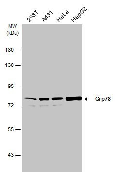
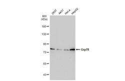
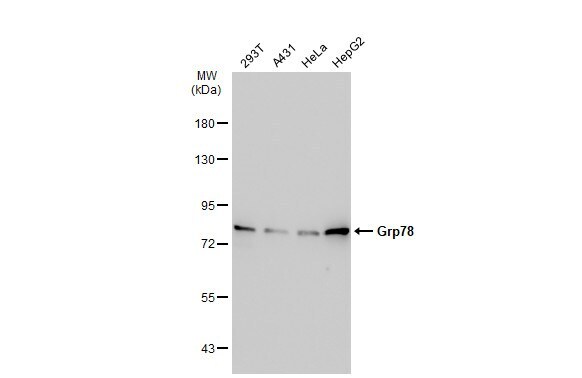
- Western blot analysis of GRP78/HSPA5 using A) 30 µg 293T whole cell lysate (B) 30 µg A431 whole cell lysate (C) 30 µg HeLa whole cell lysate and D) 30 µg HepG2 whole cell lysate. Samples were loaded onto a 7.5% SDS-PAGE gel and probed with a GRP78/HSPA5 polyclonal antibody (Product # PA5-29705) at a dilution of 1:1000.
- Submitted by
- Invitrogen Antibodies (provider)
- Main image
- Experimental details
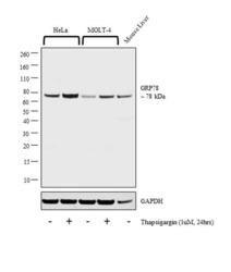
- Western blot analysis was performed on whole cell extract (30 µg lysate) of HeLa (Lane 1), HeLa treated with Thapsigargin (1uM, 24 hrs) (Lane 2), MOLT-4 (Lane 3), and MOLT-4 treated with Thapsigargin (1uM, 24 hrs) (Lane 4) and tissue lysate of Mouse liver (Lane 5). The blot was probed with Anti-GRP78 Polyclonal Antibody (Product # PA5-22967, 1:2000 dilution) and detected by chemiluminescence using Goat anti-Rabbit IgG (H+L) Superclonal™ Secondary Antibody, HRP conjugate (Product # A27036, 0.25 µg/ml, 1:4000 dilution). A 78 kDa band corresponding to GRP78 was observed in all cell lines and tissue tested.

- Submitted by
- Invitrogen Antibodies (provider)
- Main image
- Experimental details
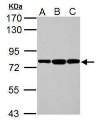
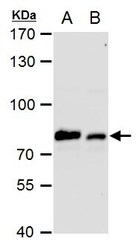
- Western Blot analysis of GRP78 was performed by separating 30 µg of various whole cell extracts by 7.5% SDS-PAGE. Proteins were transferred to a membrane and probed with a GRP78 Polyclonal Antibody (Product # PA5-29705) at a dilution of 1:10000 and a HRP-conjugated anti-rabbit IgG secondary antibody.
- Submitted by
- Invitrogen Antibodies (provider)
- Main image
- Experimental details
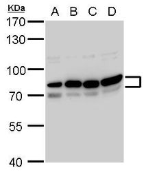
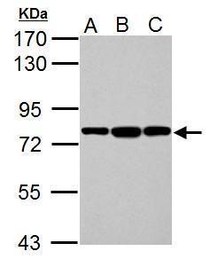
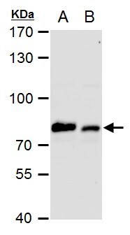
- Western Blot using GRP78 Polyclonal Antibody (Product # PA5-29705). Sample (30 µg of whole cell lysate). Lane A: NIH-3T3. Lane B: JC. Lane C: BCL-1. 7.5% SDS PAGE. GRP78 Polyclonal Antibody (Product # PA5-29705) diluted at 1:10,000. The HRP-conjugated anti-rabbit IgG antibody was used to detect the primary antibody.
- Submitted by
- Invitrogen Antibodies (provider)
- Main image
- Experimental details
- Western Blot using GRP78 Polyclonal Antibody (Product # PA5-29705). Various whole cell extracts (30 µg) were separated by 7.5% SDS-PAGE, and the membrane was blotted with GRP78 Polyclonal Antibody (Product # PA5-29705) diluted at 1:10,000. The HRP-conjugated anti-rabbit IgG antibody was used to detect the primary antibody.
- Submitted by
- Invitrogen Antibodies (provider)
- Main image
- Experimental details
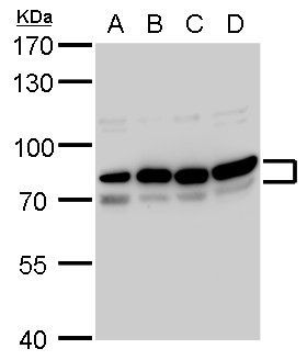
- GRP78 Polyclonal Antibody detects Grp78 protein by western blot analysis. A. 30 µg PC-12 whole cell extract. B. 30 µg Rat2 whole cell extract.7.5% SDS-PAGE. GRP78 Polyclonal Antibody (Product # PA5-29705) dilution: 1:10,000. The HRP-conjugated anti-rabbit IgG antibody was used to detect the primary antibody.
Supportive validation
- Submitted by
- Invitrogen Antibodies (provider)
- Main image
- Experimental details
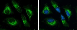
- Immunofluorescent analysis of GRP78/HSPA5 showing staining in the cytoplasm of A431 cells. A431 cells were fixed in 4% paraformaldehyde at RT for 15 min and stained using a GRP78/HSPA5 polyclonal antibody (Product # PA5-29705) diluted at 1:500. Blue: Hoechst 33342 staining.
- Submitted by
- Invitrogen Antibodies (provider)
- Main image
- Experimental details
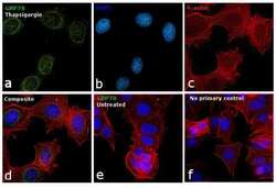
- Immunofluorescence analysis of GRP78 was performed using 70% confluent log phase MCF7 cells treated with Thapsigargin (1 µM, 24 hrs). The cells were fixed with 4% paraformaldehyde for 10 minutes, permeabilized with 0.1% Triton™ X-100 for 15 minutes, and blocked with 1% BSA for 1 hour at room temperature. The cells were labeled with GRP78 Polyclonal Antibody (Product # PA5-29705) at 5 µg/mL in 0.1% BSA, incubated at 4 degree Celsius overnight and then labeled with Goat anti-Rabbit IgG (H+L) Superclonal™ Secondary Antibody, Alexa Fluor® 488 conjugate (Product # A27034) at a dilution of 1:2000 for 45 minutes at room temperature (Panel a: green). Nuclei (Panel b: blue) were stained with ProLong™ Diamond Antifade Mountant with DAPI (Product # P36962). F-actin (Panel c: red) was stained with Rhodamine Phalloidin (Product # R415, 1:300). Panel d represents the merged image showing endoplasmic reticulum and mitochondrial localization upon treatment. Panel e represents the control untreated cells with very weak signal. Panel f represents control cells with no primary antibody to assess background. The images were captured at 60X magnification.

- Submitted by
- Invitrogen Antibodies (provider)
- Main image
- Experimental details
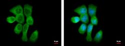
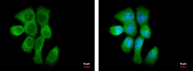
- Immunocytochemistry-Immunofluorescence analysis of GRP78 was performed in HeLa cells fixed in ice-cold MeOH for 5 min. Green: GRP78 Polyclonal Antibody (Product # PA5-29705) diluted at 1:500. Blue: Hoechst 33342 staining.

- Submitted by
- Invitrogen Antibodies (provider)
- Main image
- Experimental details
- Immunocytochemistry-Immunofluorescence analysis of GRP78 was performed in HeLa cells fixed in ice-cold MeOH for 5 min. Green: GRP78 Polyclonal Antibody (Product # PA5-29705) diluted at 1:500. Blue: Hoechst 33342 staining. Scale bar = 10 µm.

- Submitted by
- Invitrogen Antibodies (provider)
- Main image
- Experimental details
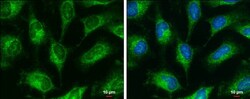
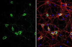
- GRP78 Polyclonal Antibody detects Grp78 protein by immunofluorescent analysis. Sample: DIV10 rat E18 primary cortical neuron cells were fixed in 4% paraformaldehyde at RT for 15 min. Green: Grp78 stained by GRP78 Polyclonal Antibody (Product # PA5-29705) diluted at 1:500. Red: Tau, stained by Tau antibody [GT287] diluted at 1:500. Blue: Fluoroshield with DAPI .

Supportive validation
- Submitted by
- Invitrogen Antibodies (provider)
- Main image
- Experimental details
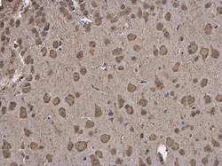
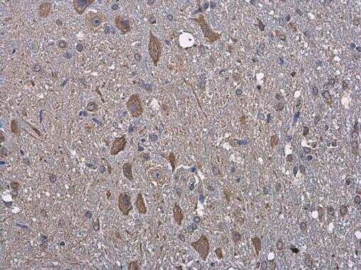
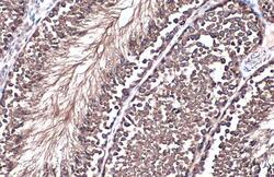
- Immunohistochemistry (Paraffin) analysis of GRP78 was performed in paraffin-embedded mouse brain tissue using GRP78 Polyclonal Antibody (Product # PA5-29705) at a dilution of 1:250.
- Submitted by
- Invitrogen Antibodies (provider)
- Main image
- Experimental details
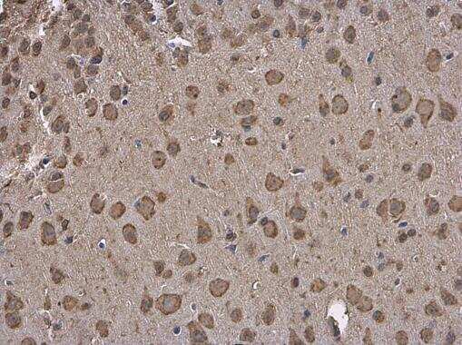
- Immunohistochemistry (Paraffin) analysis of GRP78 was performed in paraffin-embedded rat brain tissue using GRP78 Polyclonal Antibody (Product # PA5-29705) at a dilution of 1:250.
- Submitted by
- Invitrogen Antibodies (provider)
- Main image

- Experimental details
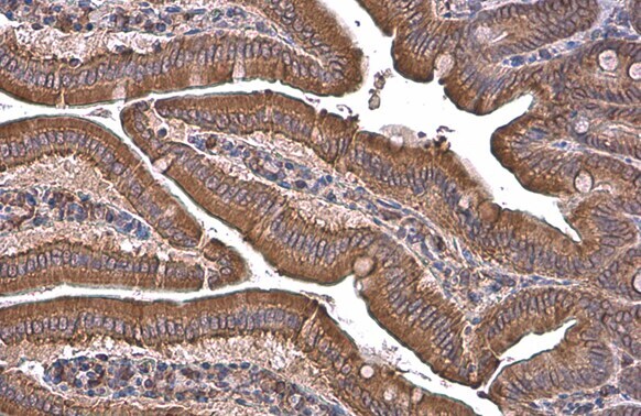
- Immunohistochemistry (Paraffin) analysis of GRP78 was performed in paraffin-embedded mouse intestine tissue using GRP78 Polyclonal Antibody (Product # PA5-29705) at a dilution of 1:500. Antigen Retrieval: Citrate buffer, pH 6.0, 15 min.
- Submitted by
- Invitrogen Antibodies (provider)
- Main image

- Experimental details
- Immunohistochemistry (Paraffin) analysis of GRP78 was performed in paraffin-embedded rat duodenum tissue using GRP78 Polyclonal Antibody (Product # PA5-29705) at a dilution of 1:500. Antigen Retrieval: Citrate buffer, pH 6.0, 15 min.
- Submitted by
- Invitrogen Antibodies (provider)
- Main image
- Experimental details
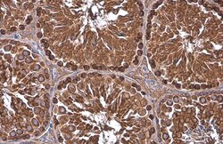
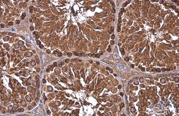
- Immunohistochemistry (Paraffin) analysis of GRP78 was performed in paraffin-embedded mouse testis tissue using GRP78 Polyclonal Antibody (Product # PA5-29705) at a dilution of 1:500. Antigen Retrieval: Citrate buffer, pH 6.0, 15 min.
- Submitted by
- Invitrogen Antibodies (provider)
- Main image

- Experimental details
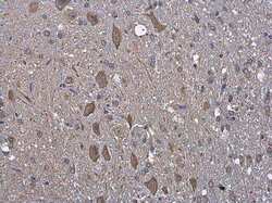
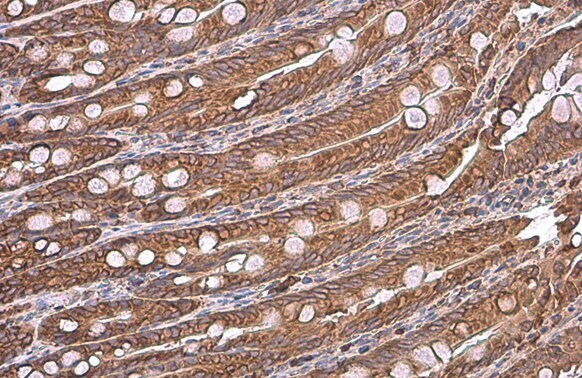
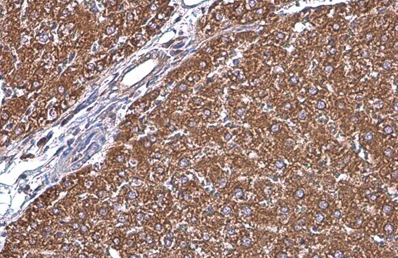
- Immunohistochemistry (Paraffin) analysis of GRP78 was performed in paraffin-embedded rat liver tissue using GRP78 Polyclonal Antibody (Product # PA5-29705) at a dilution of 1:500. Antigen Retrieval: Citrate buffer, pH 6.0, 15 min.
- Submitted by
- Invitrogen Antibodies (provider)
- Main image
- Experimental details
- GRP78 Polyclonal Antibody detects Grp78 protein at cytoplasm by immunohistochemical analysis. Sample: Paraffin-embedded mouse testis. Grp78 stained by GRP78 Polyclonal Antibody (Product # PA5-29705) diluted at 1:500. Antigen Retrieval: Citrate buffer, pH 6.0, 15 min.
