 Explore
 Validate
 Learn
Learn Western blot
Antibody data
- Antibody Data
- Antigen structure
- References [3]
- Comments [0]
- Validations
- Western blot [2]
- Immunocytochemistry [2]
- Other assay [1]
Submit
Validation data
Reference
Comment
Report error
- Product number
- PA5-16327 - Provider product page

- Provider
- Invitrogen Antibodies
- Product name
- HSP90 beta Polyclonal Antibody
- Antibody type
- Polyclonal
- Antigen
- Synthetic peptide
- Description
- PA5-16327 targets Heat Shock Protein 90 beta in IP, IF/ICC, and WB applications and shows reactivity with mouse, Rat, and Human samples.
- Concentration
- 1 mg/mL
Submitted references Functional complexity of the axonal growth cone: a proteomic analysis.
Hectd1 regulates intracellular localization and secretion of Hsp90 to control cellular behavior of the cranial mesenchyme.
Differential proteome and transcriptome analysis of porcine skeletal muscle during development.
Estrada-Bernal A, Sanford SD, Sosa LJ, Simon GC, Hansen KC, Pfenninger KH
PloS one 2012;7(2):e31858
PloS one 2012;7(2):e31858
Hectd1 regulates intracellular localization and secretion of Hsp90 to control cellular behavior of the cranial mesenchyme.
Sarkar AA, Zohn IE
The Journal of cell biology 2012 Mar 19;196(6):789-800
The Journal of cell biology 2012 Mar 19;196(6):789-800
Differential proteome and transcriptome analysis of porcine skeletal muscle during development.
Xu Y, Qian H, Feng X, Xiong Y, Lei M, Ren Z, Zuo B, Xu D, Ma Y, Yuan H
Journal of proteomics 2012 Apr 3;75(7):2093-108
Journal of proteomics 2012 Apr 3;75(7):2093-108
No comments: Submit comment
Supportive validation
- Submitted by
- Invitrogen Antibodies (provider)
- Main image
- Experimental details
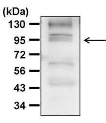
- Western blot analysis of HSP90 was performed by loading 30 µg of THP-1 whole cell lysate per well onto an SDS-PAGE gel. Proteins were transferred to a PVDF membrane and blocked with 5% non-fat dry milk in TBST for 1 hour at room temperature. The membrane was probed with an Hsp90 polyclonal antibody (Product # PA5-16327) at a dilution of 1:1000 overnight at 4°C, washed in TBST, and probed with an HRP-conjugated goat anti-rabbit IgG secondary antibody at a dilution of 1:40,000 for 1 hour at room temperature. Detection was performed using ECL substrate. Data courtesy of the Innovators Program.

- Submitted by
- Invitrogen Antibodies (provider)
- Main image
- Experimental details
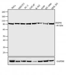
- Western blot analysis was performed on whole cell extracts (30 µg lysate) of HeLa (Lane 1), NIH/3T3 (Lane 2), PC-3 (Lane 3), LNCaP (Lane 4), K-562 (Lane 5), A549 (Lane 6), HT-1080 (Lane 7) and HEK 293 (Lane 8). The blot was probed with Rabbit Anti-HSP90 Polyclonal Antibody (Product # PA5-16327, 1 µg/mL) and detected by chemiluminescence using Goat anti-Rabbit IgG (H+L) Superclonal™ Secondary Antibody, HRP conjugate (Product # A27036, 0.4 µg/mL, 1:2500 dilution). A 90 kDa band corresponding to HSP90 was observed across the cell lines tested. Known quantity of protein samples were electrophoresed using Novex® NuPAGE® 4-12 % Bis-Tris gel (Product # NP0321BOX), XCell SureLock™ Electrophoresis System (Product # EI0002) and Novex® Sharp Pre-Stained Protein Standard (Product # LC5800). Resolved proteins were then transferred onto a nitrocellulose membrane with iBlot® 2 Dry Blotting System (Product # IB21001). The membrane was probed with the relevant primary and secondary Antibody following blocking with 5 % skimmed milk. Chemiluminescent detection was performed using Pierce™ ECL Western Blotting Substrate (Product # 32106).

Supportive validation
- Submitted by
- Invitrogen Antibodies (provider)
- Main image
- Experimental details
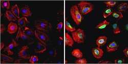
- Immunofluorescent analysis of Heat Shock Protein 90 beta (green) in HeLa cells. Formalin fixed cells were permeabilized with 0.1% Triton X-100 in TBS for 10 minutes at room temperature and blocked with 1% Blocker BSA (Product # 37525) for 15 minutes at room temperature. Cells were probed without (left panel) or with (right panel) a Heat Shock Protein 90 beta polyclonal antibody (Product # PA5-16327) at a dilution of 1:200 for at least 1 hour at room temperature, washed with PBS, and incubated with DyLight 488 goat anti-rabbit IgG secondary antibody (Product # 35552) at a dilution of 1:400 for 30 minutes at room temperature. F-Actin (red) was stained with DyLight 554 Phalloidin (Product # 21834) and nuclei (blue) were stained with Hoechst 33342 dye (Product # 62249). Images were taken on a Thermo Scientific ArrayScan or a ToxInsight Instrument at 20X magnification.

- Submitted by
- Invitrogen Antibodies (provider)
- Main image

- Experimental details
- Immunofluorescent analysis of Hsp90 beta (green) HEK293T cells. Cells fixed with 4% formaldehyde were permeabilized and blocked with 1X PBS containing 5% BSA and 0.3% Triton X-100 for 1 hour at room temperature. Cells were probed with a Hsp90 beta polyclonal antibody (Product # PA5-16327) at a dilution of 1:100 overnight at 4°C in 1X PBS containing 1% BSA and 0.3% Triton X-100, washed with 1X PBS, and incubated with a fluorophore-conjugated goat anti-rabbit IgG secondary antibody at a dilution of 1:200 for 1 hour at room temperature. Nuclei (blue) were stained with DAPI. Images were taken on a Leica DM1000 microscope at 40X magnification. Data courtesy of the Innovators Program.

Supportive validation
- Submitted by
- Invitrogen Antibodies (provider)
- Main image
- Experimental details
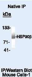
- Immunoprecipitation of Heat Shock Protein 90 beta using Heat Shock Protein 90 beta Polyclonal Antibody (Product # PA5-16327) on Native Mouse MAD109 Cells.
