 Explore
 Validate
 Learn
Learn Western blot
Antibody data
- Antibody Data
- Antigen structure
- References [1]
- Comments [0]
- Validations
- Western blot [1]
- Immunocytochemistry [1]
Submit
Validation data
Reference
Comment
Report error
- Product number
- MA5-15068 - Provider product page

- Provider
- Invitrogen Antibodies
- Product name
- IRS1 Monoclonal Antibody (K.346.8)
- Antibody type
- Monoclonal
- Antigen
- Synthetic peptide
- Description
- It is not recommended to aliquot this antibody.
- Antibody clone number
- K.346.8
- Concentration
- 84 µg/mL
Submitted references Berberine induces lipolysis in porcine adipocytes by activating the AMP‑activated protein kinase pathway.
Yang Y, Lu R, Gao F, Zhang J, Liu F
Molecular medicine reports 2020 Jun;21(6):2603-2614
Molecular medicine reports 2020 Jun;21(6):2603-2614
No comments: Submit comment
Supportive validation
- Submitted by
- Invitrogen Antibodies (provider)
- Main image

- Experimental details
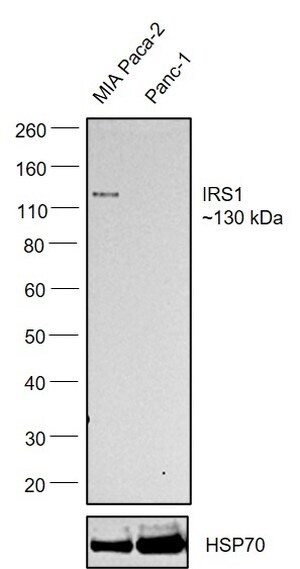
- Western blot was performed using Anti-IRS1 Monoclonal Antibody (K.346.8) (Product # MA5-15068) and a ~130kDa band corresponding to Insulin receptor substrate 1 was observed across cell lines tested . Whole cell extracts (50 µg lysate) of MIA PaCa-2 (Lane 1), PANC-1 (Lane 2) were electrophoresed using NuPAGE™ 4-12% Bis-Tris Protein Gel (Product # NP0321BOX). Resolved proteins were then transferred onto a nitrocellulose membrane (Product # IB23001) by iBlot® 2 Dry Blotting System (Product # IB21001). The blot was probed with the primary antibody (1:1000) and detected by chemiluminescence with Goat anti-Rabbit IgG (H+L) Superclonal™ Recombinant Secondary Antibody, HRP (Product # A27036,1:20000 using the iBright™ FL1500 Imaging System (Product # A44115). Chemiluminescent detection was performed using SuperSignal™ West Pico PLUS Chemiluminescent Substrate (Product # 34580).Relative expression observed between MIA Paca-2 and Panc-1.
Supportive validation
- Submitted by
- Invitrogen Antibodies (provider)
- Main image

- Experimental details
- Immunofluorescence analysis of Insulin receptor substrate 1 was performed using 70% confluent log phase MIA PaCa-2 cells. The cells were fixed with 4% paraformaldehyde for 15 minutes, permeabilized with 0.1% Triton™ X-100 for 15 minutes, and blocked with 2% BSA for 45 minutes at room temperature. The cells were labeled with IRS1 Monoclonal Antibody (K.346.8) (Product # MA5-15068) at 1:100 dilution in 0.1% BSA, incubated at 4 degree celsius overnight and then labeled with Donkey anti-Rabbit IgG (H+L) Highly Cross-Adsorbed Secondary Antibody, Alexa Fluor Plus 488 (Product # A32790), (1:2000), for 45 minutes at room temperature (Panel a: Green). Nuclei (Panel b: Blue) were stained with ProLong™ Diamond Antifade Mountant with DAPI (Product # P36962). F-actin (Panel c: Red) was stained withRhodamine Phalloidin (Product # R415, 1:300). Panel d represents the merged image showing nucleus and cytoplasm localization. Panel e represents Panc-1. Panel f represents control cells with no primary antibody to assess background. The images were captured at 60x magnification.
