 Explore
 Validate
 Learn
Learn Western blot
Antibody data
- Antibody Data
- Antigen structure
- References [0]
- Comments [0]
- Validations
- Western blot [2]
- Immunocytochemistry [2]
- Immunohistochemistry [3]
- Chromatin Immunoprecipitation [1]
Submit
Validation data
Reference
Comment
Report error
- Product number
- PA5-63349 - Provider product page

- Provider
- Invitrogen Antibodies
- Product name
- SAM68 Polyclonal Antibody
- Antibody type
- Polyclonal
- Antigen
- Recombinant full-length protein
- Description
- Immunogen sequence: ALVRGTPVRG AITRGATVTR GVPPPPTVRG APAPRARTAG IQRIPLP
- Concentration
- 0.2 mg/mL
No comments: Submit comment
Supportive validation
- Submitted by
- Invitrogen Antibodies (provider)
- Main image
- Experimental details
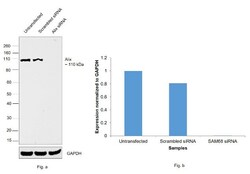
- Knockdown of SAM68 was achieved by transfecting HeLa with Alix specific siRNAs (Silencer® select Product # s20952, s20951). Western blot analysis (Fig. a) was performed using modified whole cell extracts (1% SDS) from the SAM68 knockdown cells (Lane 3), non-specific scrambled siRNA transfected cells (Lane 2) and untransfected cells (Lane 1). The blot was probed with SAM68 Polyclonal Antibody (Product # PA5-52873, 0.4 ug/ml) and Goat anti-Rabbit IgG (H+L) Superclonal™ Secondary Antibody, HRP conjugate (Product # A27036, 0.25ug/ml, 1:4000 dilution). Densitometric analysis of this Western Blot is shown in histogram (Fig. b). Decrease in signal upon siRNA mediated knock down confirms that antibody is specific to SAM68.
- Submitted by
- Invitrogen Antibodies (provider)
- Main image
- Experimental details
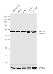
- Western blot was performed using Anti-SAM68 Polyclonal Antibody (Product # PA5-63349) and a 58 kDa band corresponding to SAM68 was observed across cell lines tested. Modified whole cell extracts (1% SDS) (30 µg lysate) of MDA-MB-231 (Lane 1), HeLa (Lane 2), A-431 (Lane 3), U-87 MG (Lane 4) and MOLT-4 (Lane 5) were electrophoresed using NuPAGE™ 10% Bis-Tris Protein Gel (Product # NP0302BOX). Resolved proteins were then transferred onto a nitrocellulose membrane (Product # IB23001) by iBlot® 2 Dry Blotting System (Product # IB21001). The blot was probed with the primary antibody (1:1000 dilution) and detected by chemiluminescence with Goat Anti-Rabbit IgG Secondary Antibody, HRP conjugate (Product # A27036, 1:4000 dilution) using the iBright FL 1000 (Product # A32752). Chemiluminescent detection was performed using Novex® ECL Chemiluminescent Substrate Reagent Kit (Product # WP20005).

Supportive validation
- Submitted by
- Invitrogen Antibodies (provider)
- Main image

- Experimental details
- Immunofluorescent staining of SAM68 in human cell line U-251 MG shows positivity in nucleus but excluded from the nucleoli. Samples were probed using a SAM68 Polyclonal Antibody (Product # PA5-63349).
- Submitted by
- Invitrogen Antibodies (provider)
- Main image

- Experimental details
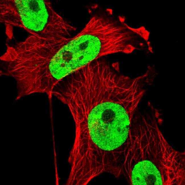
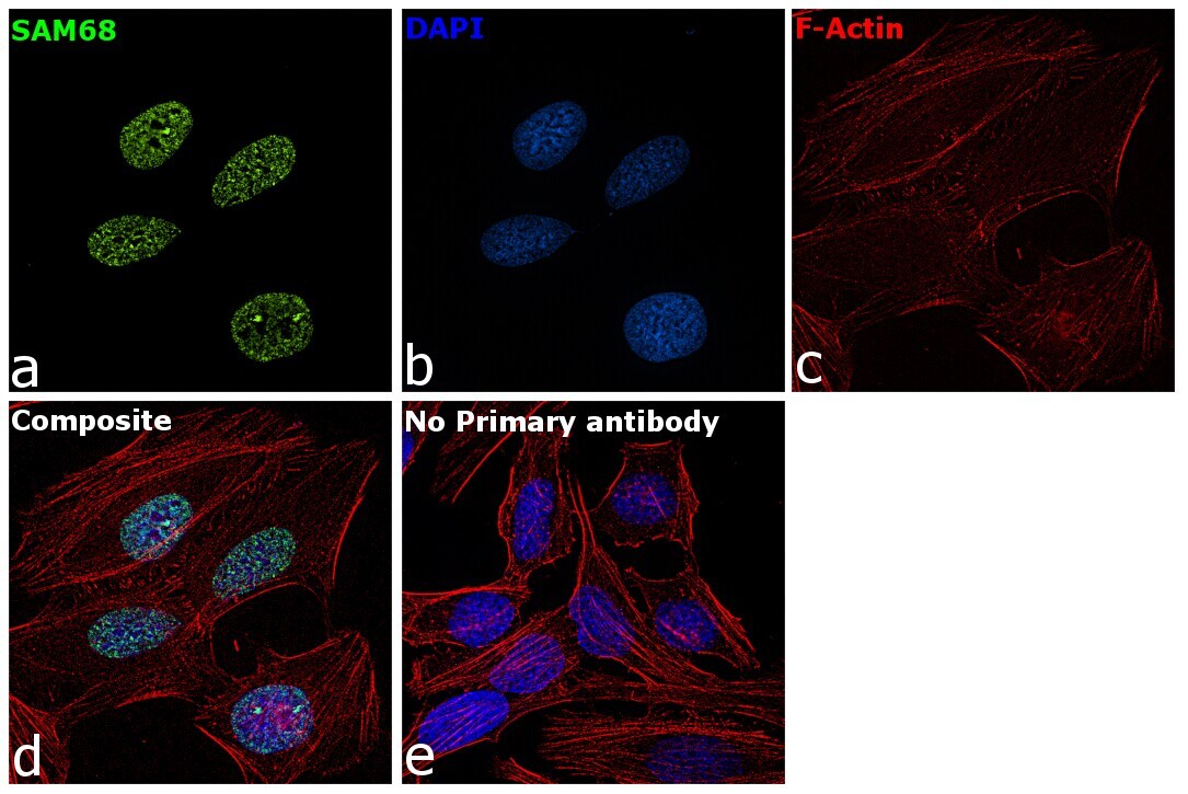
- Immunofluorescence analysis of SAM68 was performed using MCF7 cells. The cells were fixed with 4% paraformaldehyde for 10 minutes, permeabilized with 0.1% Triton™ X-100 for 15 minutes, and blocked with 2% BSA for 1 hour at room temperature. The cells were labeled with SAM68 Polyclonal Antibody (Product # PA5-63349) at 2 µg/mL in 0.1% BSA and incubated overnight at 4 degree and then labeled with Goat anti-Rabbit IgG (H+L) Superclonal™ Secondary Antibody, Alexa Fluor® 488 conjugate (Product # A27034) at a dilution of 1:2000 for 45 minutes at room temperature (Panel a: green). Nuclei (Panel b: blue) were stained with ProLong™ Diamond Antifade Mountant with DAPI (Product # P36962). F-actin (Panel c: red) was stained with Rhodamine Phalloidin (Product # R415, 1:300). Panel d represents the composite image showing nuclear localization of SAM68. Panel e represents control cells with no primary antibody to assess background. The images were captured at 60X magnification.
Supportive validation
- Submitted by
- Invitrogen Antibodies (provider)
- Main image
- Experimental details
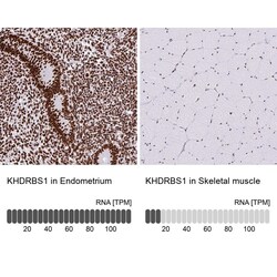
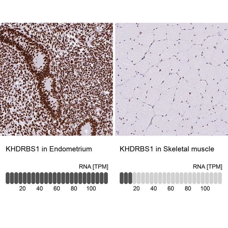
- Immunohistochemical staining of SAM68 in human endometrium and skeletal muscle tissues using SAM68 Polyclonal Antibody (Product # PA5-63349). Corresponding KHDRBS1 RNA-seq data are presented for the same tissues.
- Submitted by
- Invitrogen Antibodies (provider)
- Main image
- Experimental details
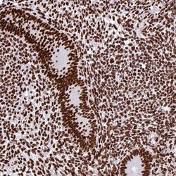
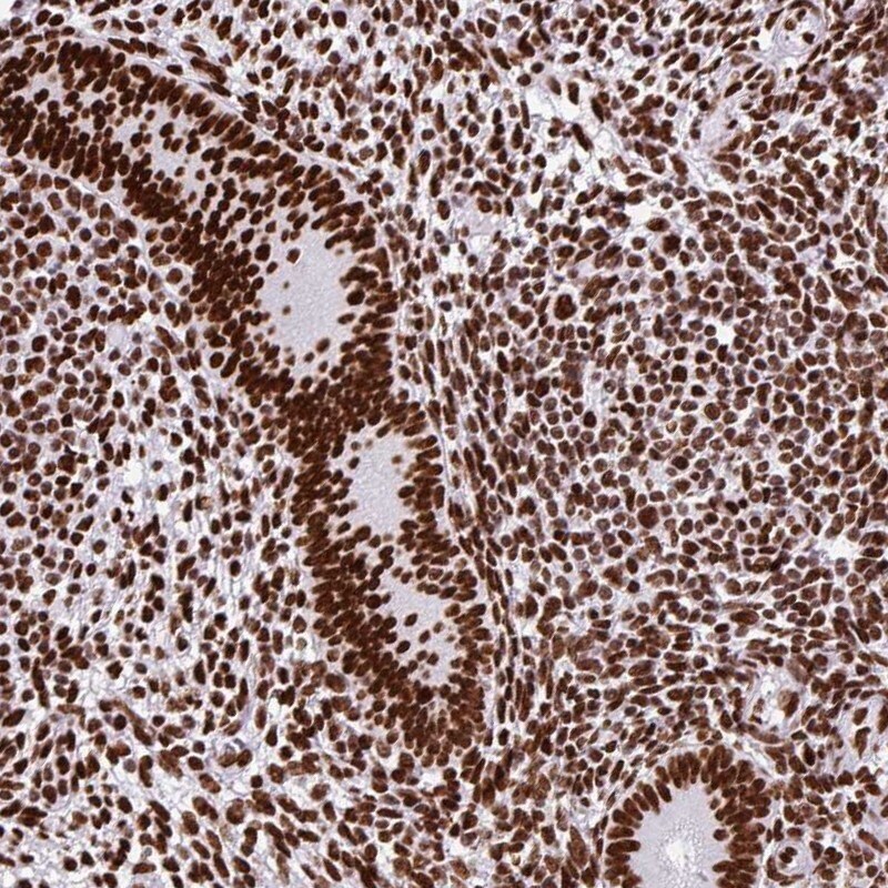
- Immunohistochemical staining of SAM68 in human endometrium using SAM68 Polyclonal Antibody (Product # PA5-63349) shows high expression.
- Submitted by
- Invitrogen Antibodies (provider)
- Main image
- Experimental details
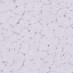
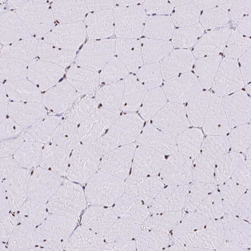
- Immunohistochemical staining of SAM68 in human skeletal muscle using SAM68 Polyclonal Antibody (Product # PA5-63349) shows low expression as expected.
Supportive validation
- Submitted by
- Invitrogen Antibodies (provider)
- Main image
- Experimental details
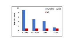
- Chromatin Immunoprecipitation (ChIP) assay of endogenous SAM68 protein using Anti-SAM68 Antibody: ChIP was performed using Anti-SAM68 Rabbit Polyclonal Antibody (Product # PA5-63349, 5 µg) on sheared chromatin from HeLa cells using the MAGnify ChIP System kit (Product # 49-2024). Normal Rabbit IgG was used as a negative IP control. The purified DNA was analyzed by qPCR using primers binding to GAPDH, NeuroD1 and cMYC transcriptional start sites and SAT alpha satellite repeats. Data is presented as fold enrichment of the antibody signal versus the negative control IgG using the comparative CT method.
