 Explore
 Validate
 Learn
Learn Western blot
Antibody data
- Antibody Data
- Antigen structure
- References [3]
- Comments [0]
- Validations
- Western blot [3]
- Other assay [2]
Submit
Validation data
Reference
Comment
Report error
- Product number
- PA1-16948 - Provider product page

- Provider
- Invitrogen Antibodies
- Product name
- VEGF Polyclonal Antibody
- Antibody type
- Polyclonal
- Antigen
- Synthetic peptide
- Description
- In Western blot, a band is seen at ~42 kDa with a chimera transfected lysate representing the homodimer VEGFA.
- Concentration
- 1 mg/mL
Submitted references PKCε Activation Restores Loss of PKCε, Manganese Superoxide Dismutase, Vascular Endothelial Growth Factor, and Microvessels in Aged and Alzheimer's Disease Hippocampus.
Uptake of Ranibizumab but Not Bevacizumab into Uveal Melanoma Cells Correlates with a Sustained Decline in VEGF-A Levels and Metastatic Activities.
Retinoic acid induces white adipose tissue browning by increasing adipose vascularity and inducing beige adipogenesis of PDGFRα(+) adipose progenitors.
Millien G, Wang H, Zhang Z, Alkon DL, Hongpaisan J
Frontiers in aging neuroscience 2022;14:836634
Frontiers in aging neuroscience 2022;14:836634
Uptake of Ranibizumab but Not Bevacizumab into Uveal Melanoma Cells Correlates with a Sustained Decline in VEGF-A Levels and Metastatic Activities.
Tura A, Pawlik VE, Rudolf M, Ernesti JS, Stutzer JN, Grisanti S, Ranjbar M
Cancers 2019 Jun 21;11(6)
Cancers 2019 Jun 21;11(6)
Retinoic acid induces white adipose tissue browning by increasing adipose vascularity and inducing beige adipogenesis of PDGFRα(+) adipose progenitors.
Wang B, Fu X, Liang X, Deavila JM, Wang Z, Zhao L, Tian Q, Zhao J, Gomez NA, Trombetta SC, Zhu MJ, Du M
Cell discovery 2017;3:17036
Cell discovery 2017;3:17036
No comments: Submit comment
Supportive validation
- Submitted by
- Invitrogen Antibodies (provider)
- Main image

- Experimental details
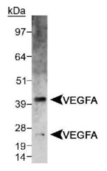
- Detection of VEGFA doublet in CSF-IR/VEGFA chimera transfected lysate using Product # PA1-16948. ECL 10 second exposure.

- Submitted by
- Invitrogen Antibodies (provider)
- Main image
- Experimental details
- Western blot analysis of VEGF in CSF-IR/VEGFA chimera transfected lysate. Sample was incubated in VEGF polyclonal antibody (Product # PA1-16948).

- Submitted by
- Invitrogen Antibodies (provider)
- Main image
- Experimental details
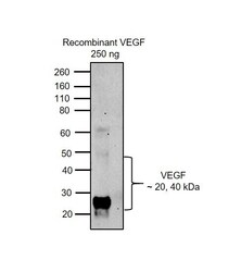
- Western blot was performed using Anti-VEGF Polyclonal Antibody (Product # PA1-16948) and 20 and 40 kDa bands corresponding to VEGF were observed in recombinant VEGF protein. Recombinant VEGF protein (250 ng) was electrophoresed using NuPAGE™ 4-12% Bis-Tris Protein Gel (Product # NP0321BOX). Resolved proteins were then transferred onto a Nitrocellulose membrane (Product # IB23002) by iBlot® 2 Dry Blotting System (Product # IB21001). The blot was probed with the primary antibody (1 µg/mL) and detected by chemiluminescence with Goat anti-Rabbit IgG (H+L) Superclonal™ Recombinant Secondary Antibody, HRP (Product # A27036, 1:4000) using the iBright FL 1000 (Product # A32752). Chemiluminescent detection was performed using Novex® ECL Reagent Kit (Product # WP20005).

Supportive validation
- Submitted by
- Invitrogen Antibodies (provider)
- Main image
- Experimental details
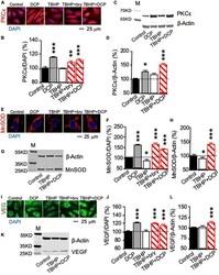
- FIGURE 2 PKCepsilon activation increases PKCepsilon, MnSOD, and VEGF protein expression in cultured human brain microvascular endothelial cells (HBMEC) treated with tert-butyl hydroperoxide (TBHP). Cultured cells were treated with 500 muM TBHP for 1 h and incubated in new culture medium with or without the PKCepsilon activators bryostatin (bry, 25 nM) and DCPLA-ME (DCP, 100 nM) for 3 days. (A,B,E,F,I,J) Immunohistochemistry imaged with confocal microscopy and (C,D,G,H,K,L) western blot analysis of (A-D) PKCepsilon, (E-H) MnSOD, and (I-L) VEGF. M, molecular weight marker. Data are represented as mean +- SEM, * p < 0.05; ** p < 0.01; *** p < 0.001; one-way ANOVA and post hoc Tukey's multiple comparison test ( n = 59-134 MnSOD-immunostained cells or 451-907 PKCepsilon or VEGF-immunostained cells from 3 to 4 cultures/group or t-test (n = 3 cultures/western blot group).

- Submitted by
- Invitrogen Antibodies (provider)
- Main image
- Experimental details
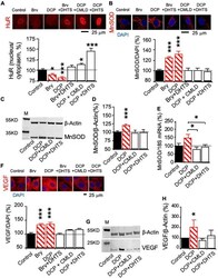
- FIGURE 4 The mRNA-stabilizing protein HuR involved in PKCepsilon-activated MnSOD and VEGF expression in human brain microvascular endothelial cells (HBMEC). HBMEC cells were treated with the HuR inhibitor CMLD-2 (35 muM) or dihydrotanshinone-I (DHTS, 10 muM) for 30 min before and during the 3-day incubation in the presence of the PKCepsilon activator bryostatin (25 nM) or DCPLA-ME (100 nM). (A) Immunohistochemistry of HuR was used to study nuclear export of the HuR protein. (B) Immunohistochemistry and (C,D) western blot analysis of MnSOD protein expression. (E) Quantitative PCR (qPCR) of MnSOD mRNA expression. (F) Immunohistochemistry and (G,H) western blot analysis of VEGF protein expression. M, molecular weight marker. Data are represented as mean +- SEM, * p < 0.05; *** p < 0.001; one-way ANOVA and post hoc Tukey's multiple comparison test ( n = 59-134 MnSOD-immunostained cells or 451-907 PKCepsilon or VEGF-immunostained cells from 3 to 4 cultures/group or t -test ( n = 3 cultures/western blot group).
