Explore
 Validate
 Learn
Learn Western blot
 Immunoprecipitation
Antibody data
- Antibody Data
- Antigen structure
- References [1]
- Comments [0]
- Validations
- Western blot [2]
- Immunohistochemistry [1]
Submit
Validation data
Reference
Comment
Report error
- Product number
- NB100-56185 - Provider product page

- Provider
- Novus Biologicals
- Proper citation
- Novus Cat#NB100-56185, RRID:AB_2272889
- Product name
- Rabbit Polyclonal XIAP Antibody
- Antibody type
- Polyclonal
- Description
- Unpurified. The antibody recognizes epitopes in the BIR2 domain of XIAP. Therefore it can recognize full-length XIAP and XIAP cleavage fragments containing the BIR2 domain. However, XIAP cleavage fragments may be biologically unstable, and therefore cleavage fragments may be difficult to detect.
- Reactivity
- Human, Mouse, Rat
- Host
- Rabbit
- Isotype
- IgG
- Vial size
- 0.05 ml
- Storage
- Store at 4C short term. Aliquot and store at -20C long term. Avoid freeze-thaw cycles.
Submitted references Phosphorylation of XIAP at threonine 180 controls its activity in Wnt signaling.
Ng VH, Hang BI, Sawyer LM, Neitzel LR, Crispi EE, Rose KL, Popay TM, Zhong A, Lee LA, Tansey WP, Huppert S, Lee E
Journal of cell science 2018 May 22;131(10)
Journal of cell science 2018 May 22;131(10)
No comments: Submit comment
Supportive validation
- Submitted by
- Novus Biologicals (provider)
- Main image
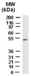
- Experimental details
- Western Blot: XIAP Antibody [NB100-56185] - Analysis of XIAP using in Human Embryonic Kidney 293 cells using this antibody.

- Submitted by
- Novus Biologicals (provider)
- Main image
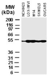
- Experimental details
- Western Blot: XIAP Antibody [NB100-56185] - Analysis of XIAP in various Tumor Cell lines using this antibody at 1:2000.

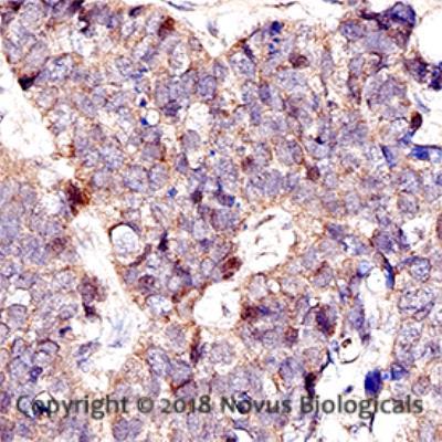
Supportive validation
- Submitted by
- Novus Biologicals (provider)
- Main image

- Experimental details
- Immunohistochemistry-Paraffin: XIAP Antibody [NB100-56185] - IHC analysis of a formalin fixed paraffin-embedded (FFPE) human breast cancer using a 1:200 dilution of of XIAP antibody on a Bond Rx autostainer (Leica Biosystems). The assay involved 20 minutes of heat induced antigen retrieval (HIER) using 10mM sodium citrate buffer (pH 6.0) and endogenous peroxidase quenching with peroxide block. The sections were incubated with primary antibody for 30 minutes and Bond Polymer Refine Detection (Leica Biosystems) with DAB was used for signal development followed by counterstaining with hematoxylin. Whole slide scanning and capturing of representative images (20X) was performed using Aperio AT2 (Leica Biosystems). Nuclear and cytoplasmic staining was observed. Staining was performed by Histowiz.