Explore
 Validate
 Learn
Learn Western blot
Antibody data
- Antibody Data
- Antigen structure
- References [7]
- Comments [0]
- Validations
- Western blot [2]
- Immunohistochemistry [3]
Submit
Validation data
Reference
Comment
Report error
- Product number
- MA3-944 - Provider product page

- Provider
- Invitrogen Antibodies
- Product name
- Calpastatin Monoclonal Antibody (1F7E3D10)
- Antibody type
- Monoclonal
- Antigen
- Purifed from natural sources
- Description
- MA3-944 detects calpastatin from bovine, human, pig and rat tissues and cells. This antibody does not cross-react with calpains or calmodulin.
- Antibody clone number
- 1F7E3D10
- Concentration
- Conc. Not Determined
Submitted references Isolation and characterization of mu-calpain, m-calpain, and calpastatin from postmortem muscle. I. Initial steps.
Purification and characterization of calpain and calpastatin from rainbow trout, Oncorhynchus mykiss.
The calpain system in human placenta.
The calpain system in human placenta.
Changes in the calpains and calpastatin during postmortem storage of bovine muscle.
Changes in the calpains and calpastatin during postmortem storage of bovine muscle.
A comparison of the intracellular distribution of mu-calpain, m-calpain, and calpastatin in proliferating human A431 cells.
Camou JP, Mares SW, Marchello JA, Vazquez R, Taylor M, Thompson VF, Goll DE
Journal of animal science 2007 Dec;85(12):3400-14
Journal of animal science 2007 Dec;85(12):3400-14
Purification and characterization of calpain and calpastatin from rainbow trout, Oncorhynchus mykiss.
Saito M, Li H, Thompson VF, Kunisaki N, Goll DE
Comparative biochemistry and physiology. Part B, Biochemistry & molecular biology 2007 Apr;146(4):445-55
Comparative biochemistry and physiology. Part B, Biochemistry & molecular biology 2007 Apr;146(4):445-55
The calpain system in human placenta.
Thompson VF, Saldaña S, Cong J, Luedke DM, Goll DE
Life sciences 2002 Apr 21;70(21):2493-508
Life sciences 2002 Apr 21;70(21):2493-508
The calpain system in human placenta.
Thompson VF, Saldaña S, Cong J, Luedke DM, Goll DE
Life sciences 2002 Apr 21;70(21):2493-508
Life sciences 2002 Apr 21;70(21):2493-508
Changes in the calpains and calpastatin during postmortem storage of bovine muscle.
Boehm ML, Kendall TL, Thompson VF, Goll DE
Journal of animal science 1998 Sep;76(9):2415-34
Journal of animal science 1998 Sep;76(9):2415-34
Changes in the calpains and calpastatin during postmortem storage of bovine muscle.
Boehm ML, Kendall TL, Thompson VF, Goll DE
Journal of animal science 1998 Sep;76(9):2415-34
Journal of animal science 1998 Sep;76(9):2415-34
A comparison of the intracellular distribution of mu-calpain, m-calpain, and calpastatin in proliferating human A431 cells.
Lane RD, Allan DM, Mellgren RL
Experimental cell research 1992 Nov;203(1):5-16
Experimental cell research 1992 Nov;203(1):5-16
No comments: Submit comment
Supportive validation
- Submitted by
- Invitrogen Antibodies (provider)
- Main image
- Experimental details
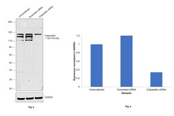
- Knockdown of Calpastatin was achieved by transfecting HeLa cells with Calpastatin specific siRNAs (Silencer® select Product # s2398, s2399). Western blot analysis (Fig. a) was performed using whole cell extracts from Calpastatin knockdown cells (Lane 3), non-specific scrambled siRNA transfected cells (Lane 2) and untransfected cells (Lane 1). The blot was probed with Calpastatin Monoclonal Antibody (1F7E3D10) (Product # MA3-944, 1:1000 dilution) and Goat anti-Mouse IgG (H+L) Superclonal™ Recombinant Secondary Antibody, HRP (Product # A28177, 1:4000 dilution). Densitometric analysis of this western blot is shown in histogram (Fig. b). Decrease in signal upon siRNA mediated knock down confirms that antibody is specific to Calpastatin.

- Submitted by
- Invitrogen Antibodies (provider)
- Main image
- Experimental details
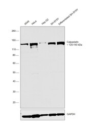
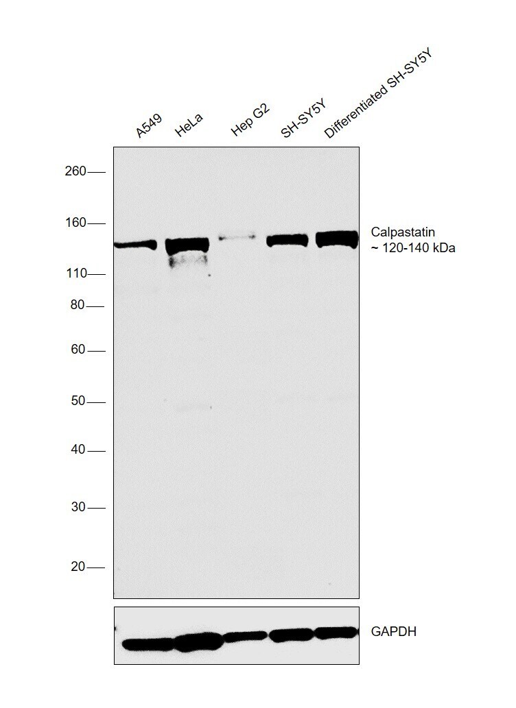
- Western blot was performed using Anti-Calpastatin Monoclonal Antibody (SP82) (Product # MA3-944) and bands at 120-140 kDa corresponding to Calpastatin was observed in all the cell lines tested and was also upregulated in differentiated SH-SY5Y when compared to SH-SY5Y. Whole cell extracts (30 µg lysate) of A549 (Lane 1), HeLa (Lane 2), Hep G2 (Lane 3), SH-SY5Y (Lane 4) and Differentiated SH-SYY (Lane 5) were electrophoresed using Novex® NuPAGE® 4-12% % Bis-Tris gel (Product # NP0322BOX). Resolved proteins were then transferred onto a nitrocellulose membrane (Product # IB23001) by iBlot® 2 Dry Blotting System (Product # IB21001). The blot was probed with the primary antibody (1:1000 dilution) and detected by chemiluminescence with Goat anti-Mouse IgG (H+L) Superclonal™ Recombinant Secondary Antibody, HRP (Product # A28177, 1:4000 dilution) using the iBright FL 1000 (Product # A32752). Chemiluminescent detection was performed using Novex® ECL Chemiluminescent Substrate Reagent Kit (Product # WP20005)..
Supportive validation
- Submitted by
- Invitrogen Antibodies (provider)
- Main image
- Experimental details
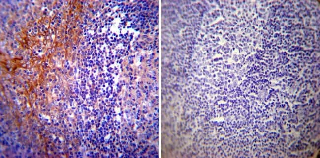
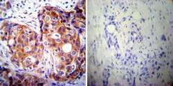
- Immunohistochemistry was performed on cancer biopsies of deparaffinized Human breast carcinoma tissues. To expose target proteins, heat induced antigen retrieval was performed using 10mM sodium citrate (pH6.0) buffer, microwaved for 8-15 minutes. Following antigen retrieval tissues were blocked in 3% BSA-PBS for 30 minutes at room temperature. Tissues were then probed at a dilution of 1:200 with a mouse monoclonal antibody recognizing Calpastatin (Product # MA3-944) or without primary antibody (negative control) overnight at 4°C in a humidified chamber. Tissues were washed extensively with PBST and endogenous peroxidase activity was quenched with a peroxidase suppressor. Detection was performed using a biotin-conjugated secondary antibody and SA-HRP, followed by colorimetric detection using DAB. Tissues were counterstained with hematoxylin and prepped for mounting.

- Submitted by
- Invitrogen Antibodies (provider)
- Main image
- Experimental details
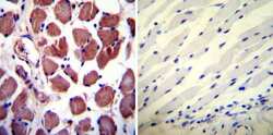
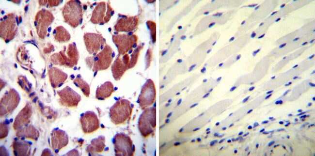
- Immunohistochemistry was performed on normal deparaffinized Human skeletal muscle tissues. To expose target proteins, heat induced antigen retrieval was performed using 10mM sodium citrate (pH6.0) buffer, microwaved for 8-15 minutes. Following antigen retrieval tissues were blocked in 3% BSA-PBS for 30 minutes at room temperature. Tissues were then probed at a dilution of 1:100 with a mouse monoclonal antibody recognizing Calpastatin (Product # MA3-944) or without primary antibody (negative control) overnight at 4°C in a humidified chamber. Tissues were washed extensively with PBST and endogenous peroxidase activity was quenched with a peroxidase suppressor. Detection was performed using a biotin-conjugated secondary antibody and SA-HRP, followed by colorimetric detection using DAB. Tissues were counterstained with hematoxylin and prepped for mounting.
- Submitted by
- Invitrogen Antibodies (provider)
- Main image

- Experimental details
- Immunohistochemistry was performed on normal deparaffinized Human tonsil tissue tissues. To expose target proteins, heat induced antigen retrieval was performed using 10mM sodium citrate (pH6.0) buffer, microwaved for 8-15 minutes. Following antigen retrieval tissues were blocked in 3% BSA-PBS for 30 minutes at room temperature. Tissues were then probed at a dilution of 1:200 with a mouse monoclonal antibody recognizing Calpastatin (Product # MA3-944) or without primary antibody (negative control) overnight at 4°C in a humidified chamber. Tissues were washed extensively with PBST and endogenous peroxidase activity was quenched with a peroxidase suppressor. Detection was performed using a biotin-conjugated secondary antibody and SA-HRP, followed by colorimetric detection using DAB. Tissues were counterstained with hematoxylin and prepped for mounting.