 Explore
 Validate
 Learn
Learn Western blot
Antibody data
- Antibody Data
- Antigen structure
- References [0]
- Comments [0]
- Validations
- Western blot [2]
- Immunocytochemistry [1]
- Immunohistochemistry [1]
Submit
Validation data
Reference
Comment
Report error
- Product number
- PA5-32722 - Provider product page

- Provider
- Invitrogen Antibodies
- Product name
- DKK1 Polyclonal Antibody
- Antibody type
- Polyclonal
- Antigen
- Synthetic peptide
- Description
- Percent identity with other species by BLAST analysis: Human, Gorilla, Monkey, Marmoset, Horse, Pig (100%); Gibbon, Mouse, Rat, Bat, Elephant (94%); Bovine, Hamster (88%); Panda, Rabbit (81%).
- Concentration
- 1 mg/mL
No comments: Submit comment
Supportive validation
- Submitted by
- Invitrogen Antibodies (provider)
- Main image
- Experimental details
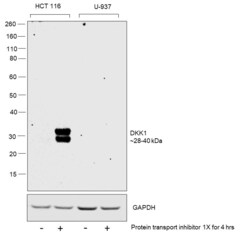
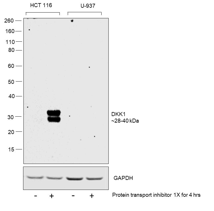
- Western blot was performed using Anti-DKK1 Polyclonal Antibody (Product # PA5-32722) and a 28-40 kDa band corresponding to DKK1 was observed only in HCT 116 on Protein transport inhibitor (1x for 4hrs) treatment but not in U-937 cells as reported in the literature. Whole-cell extracts (30 µg lysate) of HCT 116 (Lane 1), HCT 116 treated with Protein transport inhibitor (1x for 4hrs) (Lane 2), U-937 (Lane 3), U-937 treated with Protein transport inhibitor (1x for 4hrs) (Lane 4) were electrophoresed using NuPAGE™ 4-12% Bis-Tris Protein Gel (Product # NP0322BOX). Resolved proteins were then transferred onto a nitrocellulose membrane (Product # IB23002) by iBlot® 2 Dry Blotting System (Product # IB21001). The blot was probed with the primary antibody (1;1000 dilution) and detected by chemiluminescence with Goat anti-Rabbit IgG (H+L) Superclonal™ Recombinant Secondary Antibody, HRP (Product # A27036,1:20,000 dilution) using the iBright™ FL1500 Imaging System (Product # A44115). Chemiluminescent detection was performed using SuperSignal™ West Pico PLUS Chemiluminescent Substrate (Product # 34580).
- Submitted by
- Invitrogen Antibodies (provider)
- Main image
- Experimental details
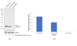
- Knockdown of DKK1 was achieved by transfecting HCT 116 cells treated with protein transport inhibitor (1x) for 4hrs) with DKK1 specific siRNAs (Silencer® select Product # S22721, S22723). Western blot analysis (Fig. a) was performed using Whole cell extracts from the DKK1 knockdown cells (lane 3), non-targeting scrambled siRNA transfected cells (lane 2), and untransfected cells (lane 1). The blot was probed with DKK1 Polyclonal Antibody (Product # PA5-32722, 1:1000 ) and Goat anti-Rabbit IgG (H+L) Superclonal™ Recombinant Secondary Antibody, HRP (Product # A27036, 1:20,000). Densitometric analysis of this western blot is shown in the histogram (Fig. b). A decrease in signal upon siRNA-mediated knockdown confirms that the antibody is specific to DKK1.

Supportive validation
- Submitted by
- Invitrogen Antibodies (provider)
- Main image

- Experimental details
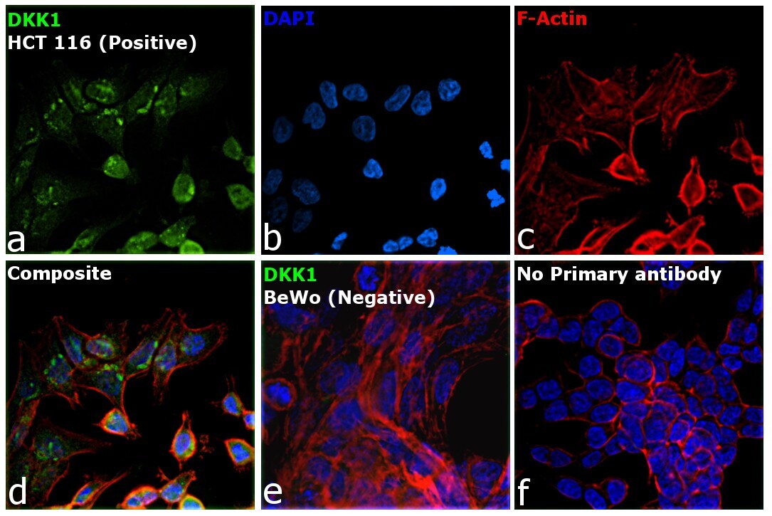
- Immunofluorescence analysis of DKK1 was performed using 70% confluent log phase HCT 116 and BeWo cells. The cells were fixed with 4% paraformaldehyde for 15 minutes, permeabilized with 0.1% Triton™ X-100 for 15 minutes, and blocked with 2% BSA for 45 minutes at room temperature. The cells were labeled with DKK1 Polyclonal Antibody (Product # PA5-32722) at 5 µg/mL in 0.1% BSA, incubated at 4 degree celsius overnight and then labeled with Donkey anti-Rabbit IgG (H+L) Highly Cross-Adsorbed Secondary Antibody, Alexa Fluor Plus 488 conjugate (Product # A32790) at a dilution of 1:2000 for 45 minutes at room temperature (Panel a: green). Nuclei (Panel b: Blue) were stained with ProLong™ Diamond Antifade Mountant with DAPI (Product # P36962). F-actin (Panel c: Red) was stained withRhodamine Phalloidin (Product # R415, 1:300). Panel d represents the merged image showing Cytoplasmic localization. Panel e represents No signal in BeWo cells. Panel f represents control cells with no primary antibody to assess background. The images were captured at 60X magnification.
Supportive validation
- Submitted by
- Invitrogen Antibodies (provider)
- Main image
- Experimental details
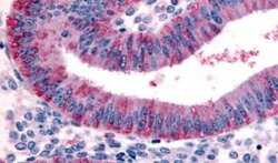
- Immunohistochemical analysis of formalin-fixed paraffin-embedded human uterus, endometrium using a DKK1 polyclonal antibody (Product # PA5-32722). Heat-induced antigen retrieval was performed prior to staining.
