Explore
 Validate
 Learn
Learn Western blot
 Immunocytochemistry
 Immunoprecipitation
Antibody data
- Antibody Data
- Antigen structure
- References [0]
- Comments [0]
- Validations
- Western blot [2]
Submit
Validation data
Reference
Comment
Report error
- Product number
- LS-C348734 - Provider product page

- Provider
- LSBio
- Product name
- Anti-S1PR1 / EDG1 / S1P1 Antibody (clone 2B9) LS-C348734
- Antibody type
- Monoclonal
- Description
- Protein A purified
- Reactivity
- Human
- Host
- Mouse
- Isotype
- IgG
- Antibody clone number
- 2B9
- Vial size
- 100µg
- Storage
- Store lyophilized at 4°C. Once reconstituted, aliquot and store at -20°C. Avoid freeze/thaw cycles. Store undiluted.
No comments: Submit comment
Supportive validation
- Submitted by
- LSBio (provider)
- Main image
- Experimental details
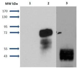
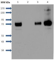
- Figure 1. Detection of S1P1 receptors in transfected cells and human umbilical vein endothelial cells (HUVEC): Lane 1: CHO-K1 cell membranes. Lane 2: S1P1-GFP CHO cell membranes. Lane 3: HUVEC membranes. CHO-K1 cells were stably transfected with S1P1-GFP construct (cell clone was a generous gift from Kevin R, Lynch). Samples were analyzed by SDS-PAGE and immunoblotted with Mouse Anti-S1PR1 Monoclonal Antibody (clone 2B9) (1/5000 w/v dilution). S1P1 receptor was fused with the green fluorescent protein (GFP) at its C-terminus. Molecular weights: GFP: 27 kDa, S1P1: 43 kDa, S1P1-GFP: 70 kDa.

- Submitted by
- LSBio (provider)
- Main image
- Experimental details
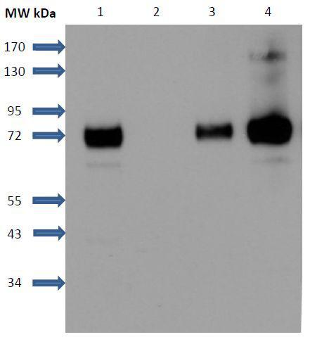
- Figure 2. Immunoprecipitation of S1P1 receptors: S1P1-GFP receptors were immunoprecipitated with a mouse monoclonal antibody anti-S1P1 after solublization of membranes with NP40 detergent and interaction with Sepharose-protein G phase. Detection of precipitated receptors was realized with a rabbit secondary anti GFP antibody coupled with HRP. Lane 1: CHO-S1P1-GFP membranes. Lane 2: Control without anti-S1P1 antibodies. Lane 3: CHO-S1P1-GFP cell membranes solubilized with NP-40. Lane 4: Immunoprecipitated sample. For control and Immunoprecipitated sample, Sepharose-protein G was boiled in Laemmli buffer for 5 min at 100°C.