Explore
 Validate
 Learn
LearnMA5-16400
antibody from Invitrogen Antibodies
Targeting: STMN1
C1orf215, FLJ32206, Lag, LAP18, OP18, PP17, PP19, PR22, SMN
 Western blot
 Flow cytometry
Antibody data
- Antibody Data
- Antigen structure
- References [0]
- Comments [0]
- Validations
- Western blot [3]
- Immunocytochemistry [1]
- Immunohistochemistry [1]
Submit
Validation data
Reference
Comment
Report error
- Product number
- MA5-16400 - Provider product page

- Provider
- Invitrogen Antibodies
- Product name
- Stathmin 1 Monoclonal Antibody (SP49)
- Antibody type
- Monoclonal
- Antigen
- Synthetic peptide
- Description
- Heat-mediated antigen retrieval is recommended prior to staining, using a 10mM citrate buffer, pH 6.0, for 10-20 minutes followed by cooling at room temperature for 20 min. Following antigen retrieval, incubate samples with primary antibody for 30 min at room temperature. A suggested positive control is tonsil or breast carcinoma.
- Reactivity
- Human, Mouse, Rat
- Host
- Rabbit
- Isotype
- IgG
- Antibody clone number
- SP49
- Vial size
- 500 µL
- Storage
- 4° C, do not freeze
No comments: Submit comment
Supportive validation
- Submitted by
- Invitrogen Antibodies (provider)
- Main image

- Experimental details
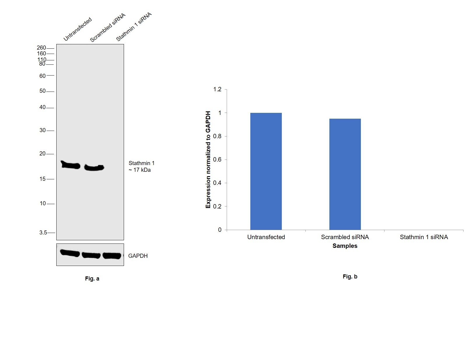
- Knockdown of Stathmin 1 was achieved by transfecting HeLa with Stathmin 1 specific siRNAs (Silencer® select Product # s8092, s8093). Western blot analysis (Fig. a) was performed using whole cell extracts from the Stathmin 1 knockdown cells (lane 3), non-targeting scrambled siRNA transfected cells (lane 2) and untransfected cells (lane 1). The blot was probed with Stathmin 1 Monoclonal Antibody (SP49) (Product # MA5-16400, 1:300 dilution ) and Goat anti-Rabbit IgG (H+L) Superclonal™ Recombinant Secondary Antibody, HRP (Product # A27036, 1:6000 dilution). Densitometric analysis of this western blot is shown in histogram (Fig. b). Decrease in signal upon siRNA mediated knock down confirms that antibody is specific to Stathmin 1.
- Submitted by
- Invitrogen Antibodies (provider)
- Main image
- Experimental details
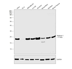
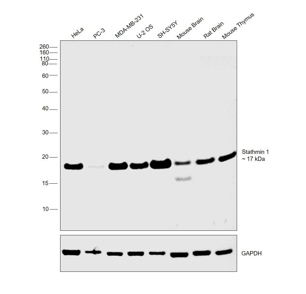
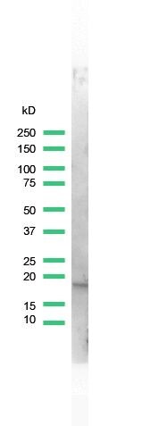
- Western blot was performed using Anti-Stathmin 1 Monoclonal Antibody (SP49) (Product # MA5-16400) and a 17kDa band corresponding to Stathmin 1 was observed across cell lines and tissue tested. Whole cell extracts (30 µg lysate) of HeLa (Lane 1), PC-3 (Lane 2), MDA-MB-231 (Lane 3), U-2 OS (Lane 4), SH-SY5Y (Lane 5), Mouse Brain (Lane 6), Rat Brain (Lane 7), Mouse Thymus (Lane 8) were electrophoresed using NuPAGE™ 12% Bis-Tris Protein Gel (Product # NP0341BOX). Resolved proteins were then transferred onto a Nitrocellulose membrane (Product # IB23001) by iBlot® 2 Dry Blotting System (Product # IB21001). The blot was probed with the primary antibody (1:300 dilution) and detected by chemiluminescence with Goat anti-Rabbit IgG (H+L) Superclonal™ Recombinant Secondary Antibody, HRP (Product # A27036, 1:6000 dilution) using the iBright FL 1000 (Product # A32752). Chemiluminescent detection was performed using Novex® ECL Chemiluminescent Substrate Reagent Kit (Product # WP20005).
- Submitted by
- Invitrogen Antibodies (provider)
- Main image
- Experimental details
- Western blot analysis of NIH3T3 Cells using anti-Stathmin Monoclonal Antibody (Product # MA5-16400). The recommened dilution for this antibody in western blot applications is 1:25.
Supportive validation
- Submitted by
- Invitrogen Antibodies (provider)
- Main image
- Experimental details
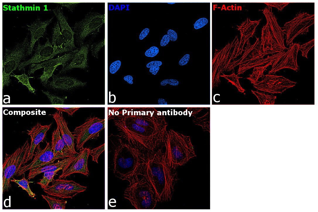
- Immunofluorescence analysis of Stathmin 1 was performed using 70% confluent log phase HeLa cells. The cells were fixed with 4% paraformaldehyde for 10 minutes, permeabilized with 0.1% Triton™ X-100 for 15 minutes, and blocked with 2% BSA for 45 minutes at room temperature. The cells were labeled with Stathmin 1 Monoclonal Antibody (SP49) (Product # MA5-16400) at 1:100 dilution in 0.1% BSA, incubated at 4 degree celsius overnight and then labeled with Donkey anti-Rabbit IgG (H+L) Highly Cross-Adsorbed Secondary Antibody, Alexa Fluor Plus 488 (Product # A32790), (1:2000 dilution), for 45 minutes at room temperature (Panel a: Green). Nuclei (Panel b:Blue) were stained with ProLong™ Diamond Antifade Mountant with DAPI (Product # P36962). F-actin (Panel c: Red) was stained with Rhodamine Phalloidin (Product # R415, 1:300). Panel d represents the merged image showing cytoplasmic localization. Panel e represents control cells with no primary antibody to assess background. The images were captured at 60X magnification.
Supportive validation
- Submitted by
- Invitrogen Antibodies (provider)
- Main image
- Experimental details
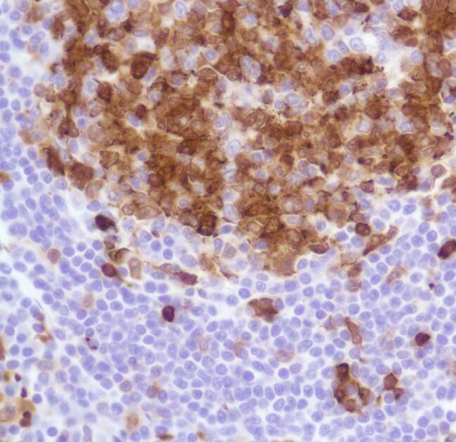
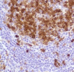
- Immunohistochemical analysis of Stathmin using anti-Stathmin Monoclonal Antibody (Product # MA5-16400) in Tonsil Tissue. The recommened dilution for this antibody in immunohistochemistry applications is 1:100.