Explore
 Validate
 Learn
LearnMA5-27123
antibody from Invitrogen Antibodies
Targeting: GNAS
GNAS1, GNASXL, GPSA, NESP, NESP55, SCG6, SgVI
 Western blot
Antibody data
- Antibody Data
- Antigen structure
- References [0]
- Comments [0]
- Validations
- Western blot [6]
- Immunohistochemistry [3]
Submit
Validation data
Reference
Comment
Report error
- Product number
- MA5-27123 - Provider product page

- Provider
- Invitrogen Antibodies
- Product name
- GNAS Monoclonal Antibody (OTI7A4)
- Antibody type
- Monoclonal
- Antigen
- Recombinant full-length protein
- Reactivity
- Human, Mouse, Rat
- Host
- Mouse
- Isotype
- IgG
- Antibody clone number
- OTI7A4
- Vial size
- 100 µL
- Concentration
- 1 mg/mL
- Storage
- -20° C, Avoid Freeze/Thaw Cycles
No comments: Submit comment
Supportive validation
- Submitted by
- Invitrogen Antibodies (provider)
- Main image

- Experimental details
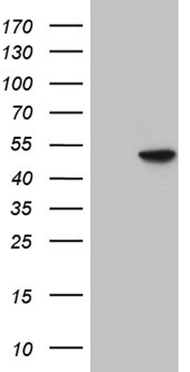
- Western blot analysis of GNAS in HEK293T cells in untransfected (Left lane) and transfected (Right lane) samples using 5 µg per lane. The samples were separated by SDS-PAGE and probed with GNAS (Product # MA5-27123) monoclonal antibody.
- Submitted by
- Invitrogen Antibodies (provider)
- Main image

- Experimental details
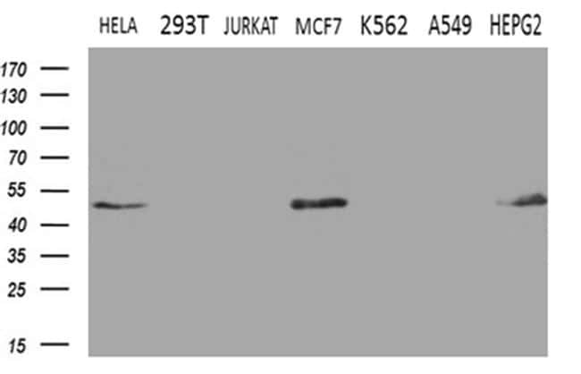
- Western blot analysis of GNAS in HeLa, 293T, Jurkat, MCF7, K562, A549, HepG2, cells using 35 µg per lane. Samples were probed with GNAS (Product # MA5-27123) monoclonal antibody at a dilution of 1:500.
- Submitted by
- Invitrogen Antibodies (provider)
- Main image
- Experimental details
- Western blot analysis of GNAS in HEK293T cells in untransfected (Left lane) and transfected (Right lane) samples using 5 µg per lane. The samples were separated by SDS-PAGE and probed with GNAS (Product # MA5-27123) monoclonal antibody.

- Submitted by
- Invitrogen Antibodies (provider)
- Main image

- Experimental details
- Western blot analysis of GNAS in HeLa, 293T, Jurkat, MCF7, K562, A549, HepG2, cells using 35 µg per lane. Samples were probed with GNAS (Product # MA5-27123) monoclonal antibody at a dilution of 1:500.

- Submitted by
- Invitrogen Antibodies (provider)
- Main image
- Experimental details
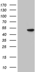
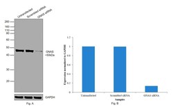
- Knockdown of GNAS was achieved by transfecting HepG2 with GNAS specific siRNAs (Silencer® select Products # s526399, s526398). Western blot analysis (Fig. a) was performed using modified whole cell extracts (1% SDS) from the GNAS knockdown cells (Lane 3), non-specific scrambled siRNA transfected cells (Lane 2) and untransfected cells (Lane 1). The blot was probed with GNAS Monoclonal Antibody (OTI7A4) (Product # MA5-27123, 1:1000 dilution)and Goat anti-Mouse IgG (H+L) Superclonal™ Recombinant Secondary Antibody, HRP (Product # A28177, 1:4000 dilution). Densitometric analysis of this western blot is shown in histogram (Fig. b). Reduction in signal upon siRNA mediated knock down confirms that antibody is specific to GNAS.

- Submitted by
- Invitrogen Antibodies (provider)
- Main image
- Experimental details
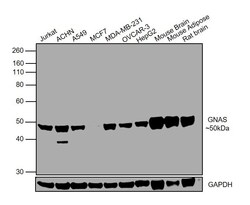
- Western blot was performed using Anti-GNAS Monoclonal Antibody (OTI7A6) (Product # MA5-27123) and a 50kDa band corresponding to GNAS was observed in all tested cell lines and tissue models. Whole cell lysates (30ug lysate) of Jurkat (Lane 1), ACHN (Lane 2), A549 (Lane 3), MCF7 (Lane 4), MDA-MB-231 (Lane 5), OVCAR-3 (Lane 6), HepG2 (Lane 7), tissue lysates (30ug lysate) of Mouse Brain (Lane 8), Mouse Adipose (Lane 9) and Rat Brain (Lane 10) were electrophoresed using NuPAGE® 10 % Bis-Tris gel (Product # NP0302BOX). Resolved proteins were then transferred onto a nitrocellulose membrane (Product # IB23001) by iBlot® 2 Dry Blotting System (Product # IB21001). The blot was probed with the primary antibody (1:1000 dilution) and detected by chemiluminescence with Goat anti-Mouse IgG (H+L) Superclonal™ Recombinant Secondary Antibody, HRP (Product # A28177, 1:4000 dilution) using the iBright FL 1000 (Product # A32752). Chemiluminescent detection was performed using Novex® ECL Chemiluminescent Substrate Reagent Kit (Product # WP20005).

Supportive validation
- Submitted by
- Invitrogen Antibodies (provider)
- Main image

- Experimental details
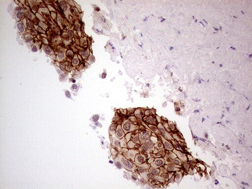
- Immunohistochemistry was performed on paraffin-embedded carcinoma of human lung tissue. To expose target proteins, heat-induced epitope retrieval by 10mM citric buffer, pH6.0, 100°C for 10min. Following antigen retrieval, tissues were probed with a GNAS monoclonal antibody (Product # MA5-27123) at a dilution of 1:500.
- Submitted by
- Invitrogen Antibodies (provider)
- Main image

- Experimental details
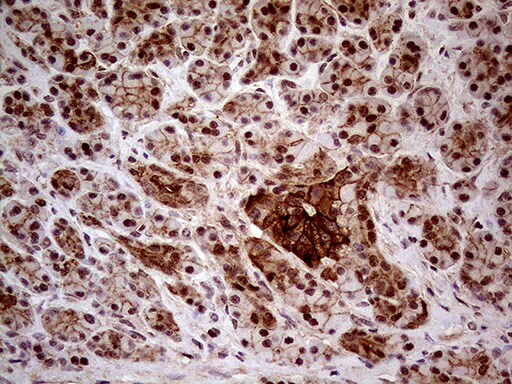
- Immunohistochemistry was performed on paraffin-embedded human pancreas tissue. To expose target proteins, heat-induced epitope retrieval by 10mM citric buffer, pH6.0, 100°C for 10min. Following antigen retrieval, tissues were probed with a GNAS monoclonal antibody (Product # MA5-27123) at a dilution of 1:500.
- Submitted by
- Invitrogen Antibodies (provider)
- Main image

- Experimental details
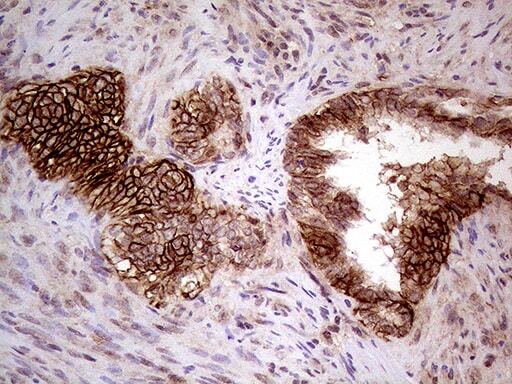
- Immunohistochemistry was performed on paraffin-embedded adenocarcinoma of human endometrium tissue. To expose target proteins, heat-induced epitope retrieval by 10mM citric buffer, pH6.0, 100°C for 10min. Following antigen retrieval, tissues were probed with a GNAS monoclonal antibody (Product # MA5-27123) at a dilution of 1:500.