Explore
 Validate
 Learn
LearnPA5-22261
antibody from Invitrogen Antibodies
Targeting: GNAS
GNAS1, GNASXL, GPSA, NESP, NESP55, SCG6, SgVI
 Western blot
Antibody data
- Antibody Data
- Antigen structure
- References [1]
- Comments [0]
- Validations
- Western blot [7]
- Immunohistochemistry [1]
Submit
Validation data
Reference
Comment
Report error
- Product number
- PA5-22261 - Provider product page

- Provider
- Invitrogen Antibodies
- Product name
- GNAS Polyclonal Antibody
- Antibody type
- Polyclonal
- Antigen
- Recombinant protein fragment
- Description
- Recommended positive controls: HepG2, Raji, mouse brain, NIH-3T3, JC, rat brain.
- Concentration
- 1.99 mg/mL
Submitted references Combined Small Cell Carcinoma of the Lung: Is It a Single Entity?
Zhao X, McCutcheon JN, Kallakury B, Chahine JJ, Pratt D, Raffeld M, Chen Y, Wang C, Giaccone G
Journal of thoracic oncology : official publication of the International Association for the Study of Lung Cancer 2018 Feb;13(2):237-245
Journal of thoracic oncology : official publication of the International Association for the Study of Lung Cancer 2018 Feb;13(2):237-245
No comments: Submit comment
Supportive validation
- Submitted by
- Invitrogen Antibodies (provider)
- Main image
- Experimental details
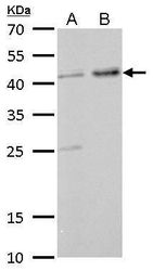
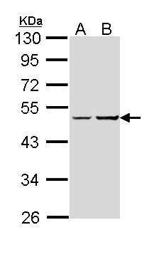
- Western blot analysis of XLalphas using A) 30 µg NIH-3T3 whole cell lysate and B) 30 µg JC whole cell lysate. Samples were loaded onto a 12% SDS-PAGE gel and probed with a XLalphas polyclonal antibody (Product # PA5-22261) at a dilution of 1:1000.
- Submitted by
- Invitrogen Antibodies (provider)
- Main image

- Experimental details
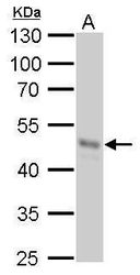
- Western blot analysis of XLalphas using 50 µg of mouse brain lysate. Samples were loaded onto a 10% SDS-PAGE gel and probed with a XLalphas polyclonal antibody (Product # PA5-22261) at a dilution of 1:1000.

- Submitted by
- Invitrogen Antibodies (provider)
- Main image
- Experimental details
- Western blot analysis of XLalphas using 50 µg rat brain lysate. Samples were loaded onto a 10% SDS-PAGE gel and probed with a XLalphas polyclonal antibody (Product # PA5-22261) at a dilution of 1:1000.

- Submitted by
- Invitrogen Antibodies (provider)
- Main image

- Experimental details
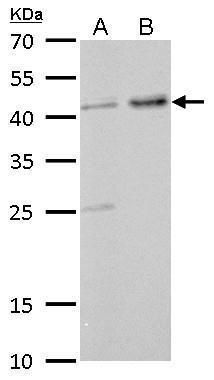
- Western blot analysis of XLalphas using 30 µg of A) HepG2 and B) Raji lysate. Samples were loaded onto a 10% SDS-PAGE gel and probed with a XLalphas polyclonal antibody (Product # PA5-22261) at a dilution of 1:1000.
- Submitted by
- Invitrogen Antibodies (provider)
- Main image
- Experimental details
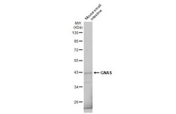
- Western Blot using GNAS Polyclonal Antibody (Product # PA5-22261). Mouse tissue extract (50 µg) was separated by 10% SDS-PAGE, and the membrane was blotted with GNAS Polyclonal Antibody (Product # PA5-22261) diluted at 1:5,000. The HRP-conjugated anti-rabbit IgG antibody was used to detect the primary antibody.

- Submitted by
- Invitrogen Antibodies (provider)
- Main image
- Experimental details
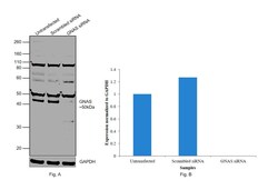
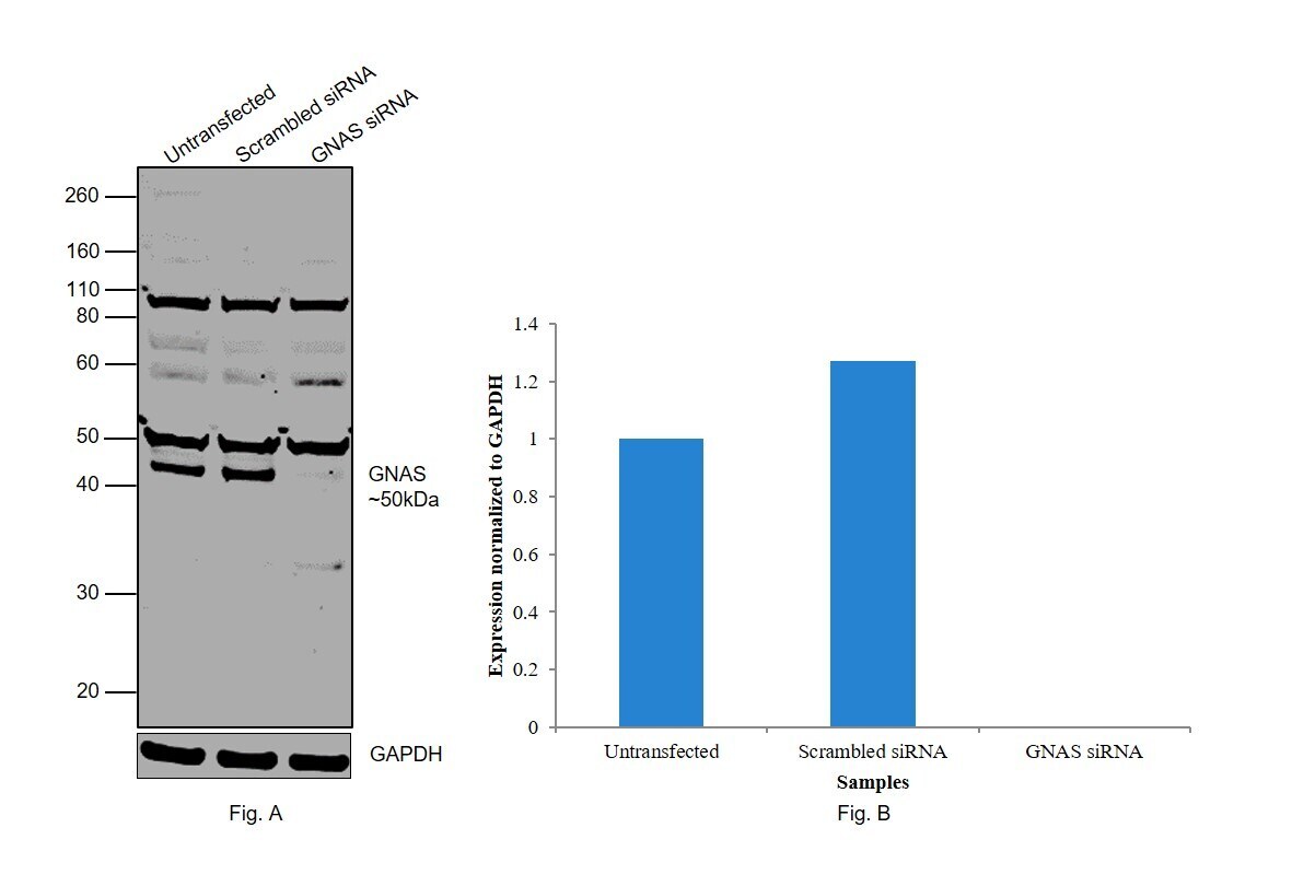
- Knockdown of GNAS was achieved by transfecting Hep G2 with GNAS specific siRNAs (Silencer® select Product # s526399, s526398). Western blot analysis (Fig. a) was performed using whole cell extracts from the GNAS knockdown cells (Lane 3), non-specific scrambled siRNA transfected cells (Lane 2) and untransfected cells (Lane 1). The blot was probed with GNAS Polyclonal Antibody (Product # PA5-22261, 1:1000 dilution) and Goat anti-Rabbit IgG (H+L) Superclonal™ Recombinant Secondary Antibody, HRP (Product # A27036, 1:4000 dilution). Densitometric analysis of this western blot is shown in histogram (Fig. b). Loss of signal upon siRNA mediated knock down confirms that antibody is specific to GNAS.
- Submitted by
- Invitrogen Antibodies (provider)
- Main image

- Experimental details
- Western blot was performed using Anti-GNAS Polyclonal Antibody (Product # PA5-22261) and a 50 kDa band corresponding to GNAS was observed in Jurkat, ACHN, MCF7, OVCAR3, Hep G2, Mouse Brain and Mouse Adipose, but not in A549, MDA-MB-231 and Rat Brain. An uncharacterized band was observed at ~52 kDa. Whole cell lysates (30 µg lysate) of Jurkat (Lane 1), ACHN (Lane 2), A549 (Lane 3), MCF7 (Lane 4), MDA-MB-231 (Lane 5), OVCAR-3 (Lane 6), Hep G2 (Lane 7), tissue Lysates (30 µg lysate) of Mouse Brain (Lane 8), Mouse Adipose (Lane 9) and Rat Brain (Lane 10) were electrophoresed using NuPAGE® 10 % Bis-Tris gel (Product # NP0302BOX). Resolved proteins were then transferred onto a nitrocellulose membrane (Product # IB23001) by iBlot® 2 Dry Blotting System (Product # IB21001). The blot was probed with the primary antibody (1:1000 dilution) and detected by chemiluminescence with Goat anti-Rabbit IgG (H+L), Superclonal™ Recombinant Secondary Antibody, HRP (Product # A27036, 1:4000 dilution) using the iBright FL 1000 (Product # A32752). Chemiluminescent detection was performed using Novex® ECL Chemiluminescent Substrate Reagent Kit (Product # WP20005).

Supportive validation
- Submitted by
- Invitrogen Antibodies (provider)
- Main image
- Experimental details
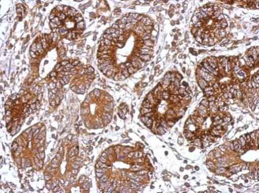
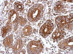
- Immunohistochemical analysis of paraffin-embedded human colon carcinoma, using GNAS (Product # PA5-22261) antibody at 1:500 dilution. Antigen Retrieval: EDTA based buffer, pH 8.0, 15 min.