Explore
 Validate
 Learn
LearnPA5-31144
antibody from Invitrogen Antibodies
Targeting: RAD51
BRCC5, FANCR, HsRad51, HsT16930, RAD51A, RECA
 Western blot
Antibody data
- Antibody Data
- Antigen structure
- References [0]
- Comments [0]
- Validations
- Western blot [4]
- Immunocytochemistry [1]
- Immunohistochemistry [2]
- Other assay [1]
Submit
Validation data
Reference
Comment
Report error
- Product number
- PA5-31144 - Provider product page

- Provider
- Invitrogen Antibodies
- Product name
- RAD51 Polyclonal Antibody
- Antibody type
- Polyclonal
- Antigen
- Recombinant protein fragment
- Description
- Recommended positive controls: 293T , NT2D, IMR32, Jurkat, HeLa, PC-12, Rat-2.
- Concentration
- 0.63 mg/mL
No comments: Submit comment
Supportive validation
- Submitted by
- Invitrogen Antibodies (provider)
- Main image

- Experimental details
- Western Blot analysis of RAD51 was performed by separating 30 µg of various whole cell extracts by 10% SDS-PAGE. Proteins were transferred to a membrane and probed with a RAD51 Polyclonal Antibody (Product # PA5-31144) at a dilution of 1:500.

- Submitted by
- Invitrogen Antibodies (provider)
- Main image

- Experimental details
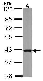
- Western Blot using RAD51 Polyclonal Antibody (Product # PA5-31144). Sample (30 µg of whole cell lysate). Lane A: JurKat. 10% SDS PAGE. RAD51 Polyclonal Antibody (Product # PA5-31144) diluted at 1:1,000.
- Submitted by
- Invitrogen Antibodies (provider)
- Main image
- Experimental details
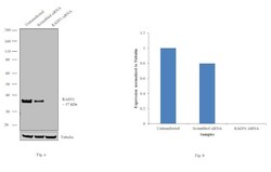
- Knockdown of RAD51 was achieved by transfecting HeLa with RAD51 specific siRNAs (Silencer® select Product # s11734, s11735). Western blot analysis (Fig. a) was performed using whole cell extracts from the RAD51 knockdown cells (lane 3), non-specific scrambled siRNA transfected cells (lane 2) and untransfected cells (lane 1). The blots were probed with RAD51 Polyclonal Antibody (Product # PA5-31144, 1:1000 dilution) and Goat anti-Rabbit IgG (H+L) Superclonal™ Secondary Antibody, HRP conjugate (Product # A27036, 0.25µg/ml, 1:4000 dilution). Densitometric analysis of this western blot is shown in histogram (Fig. b). Loss of signal upon siRNA mediated knock down confirms that antibody is specific to RAD51.

- Submitted by
- Invitrogen Antibodies (provider)
- Main image

- Experimental details
- Western blot analysis was performed on modified whole cell extracts (1% SDS) (30 µg lysate) of NIH/3T3 (Lane 1), HeLa (Lane 2), RAW 264.7 (Lane 3), Neuro-2A (Lane 4), Jurkat (Lane 5), Raji (Lane 6) and PANC-1 (Lane 7). The blot was probed with Anti-RAD51 Polyclonal Antibody (Product # PA5-31144, 1:1000 dilution) and detected by chemiluminescence using Goat anti-Rabbit IgG (H+L) Superclonal™ Secondary Antibody, HRP conjugate (Product # A27036, 0.25 µg/ml, 1:4000 dilution). Band of ~40 kDa corresponding to RAD51 was observed in cell lines tested along with additional bands at ~78 kDa and 48 kDa.

Supportive validation
- Submitted by
- Invitrogen Antibodies (provider)
- Main image
- Experimental details
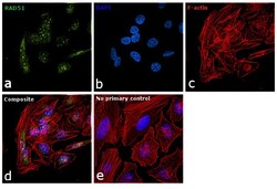
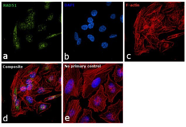
- Immunofluorescence analysis of RAD51 was performed using 70% confluent log phase HeLa cells. The cells were fixed with 4% paraformaldehyde for 10 minutes, permeabilized with 0.1% Triton™ X-100 for 15 minutes, and blocked with 1% BSA for 1 hour at room temperature. The cells were labeled with RAD51 Polyclonal Antibody (Product # PA5-31144) at 1:200 dilution in 0.1% BSA, incubated at 4 degree Celsius overnight and then labeled with Goat anti-Rabbit IgG (H+L) Superclonal™ Secondary Antibody, Alexa Fluor® 488 conjugate (Product # A27034) at a dilution of 1:2000 for 45 minutes at room temperature (Panel a: green). Nuclei (Panel b: blue) were stained with ProLong™ Diamond Antifade Mountant with DAPI (Product # P36962). F-actin (Panel c: red) was stained with Rhodamine Phalloidin (Product # R415). Panel d represents the merged image showing nuclear, cytoplasmic and perinuclear localization. Panel e represents control cells with no primary antibody to assess background. The images were captured at 60X magnification.
Supportive validation
- Submitted by
- Invitrogen Antibodies (provider)
- Main image

- Experimental details
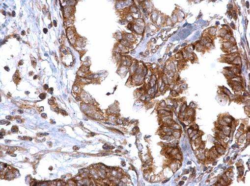
- RAD51 Polyclonal Antibody detects Rad51 protein at cytosol and nucleus on human breast carcinoma by immunohistochemical analysis. Sample: Paraffin-embedded human breast carcinoma. RAD51 Polyclonal Antibody (Product # PA5-31144) dilution: 1:500. Antigen Retrieval: EDTA based buffer, pH 8.0, 15 min.

- Submitted by
- Invitrogen Antibodies (provider)
- Main image

- Experimental details
- RAD51 Polyclonal Antibody detects Rad51 protein at cytosol on human ovarian carcinoma by immunohistochemical analysis. Sample: Paraffin-embedded human ovarian carcinoma. RAD51 Polyclonal Antibody (Product # PA5-31144) dilution: 1:500. Antigen Retrieval: EDTA based buffer, pH 8.0, 15 min.
Supportive validation
- Submitted by
- Invitrogen Antibodies (provider)
- Main image

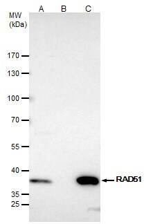
- Experimental details
- RAD51 Polyclonal Antibody immunoprecipitates Rad51 protein in IP experiments. IP samples: Jurkat whole cell extract. A. 40 µg Jurkat whole cell extract. B. Control with 4 µg of preimmune Rabbit IgG. C. Immunoprecipitation of Rad51 protein by 4 µg RAD51 Polyclonal Antibody (Product # PA5-31144). 5 % SDS-PAGE. The immunoprecipitated Rad51 protein was detected by RAD51 Polyclonal Antibody (Product # PA5-31144) diluted at 1:500.