 Explore
 Validate
 Learn
Learn Western blot
 ELISA
ELISAAntibody data
- Antibody Data
- Antigen structure
- References [9]
- Comments [0]
- Validations
- Western blot [2]
- Immunocytochemistry [1]
- Other assay [6]
Submit
Validation data
Reference
Comment
Report error
- Product number
- 37-4400 - Provider product page

- Provider
- Invitrogen Antibodies
- Product name
- EphA2 Monoclonal Antibody (1C11A12)
- Antibody type
- Monoclonal
- Antigen
- Synthetic peptide
- Reactivity
- Human, Mouse, Rat
- Host
- Mouse
- Isotype
- IgG
- Antibody clone number
- 1C11A12
- Vial size
- 100 µg
- Concentration
- 0.5 mg/mL
- Storage
- -20°C
Submitted references EPHA2 Interacts with DNA-PK(cs) in Cell Nucleus and Controls Ionizing Radiation Responses in Non-Small Cell Lung Cancer Cells.
microRNA-451a promoter methylation regulated by DNMT3B expedites bladder cancer development via the EPHA2/PI3K/AKT axis.
A novel immortalized hepatocyte-like cell line (imHC) supports in vitro liver stage development of the human malarial parasite Plasmodium vivax.
Ephrin B3 interacts with multiple EphA receptors and drives migration and invasion in non-small cell lung cancer.
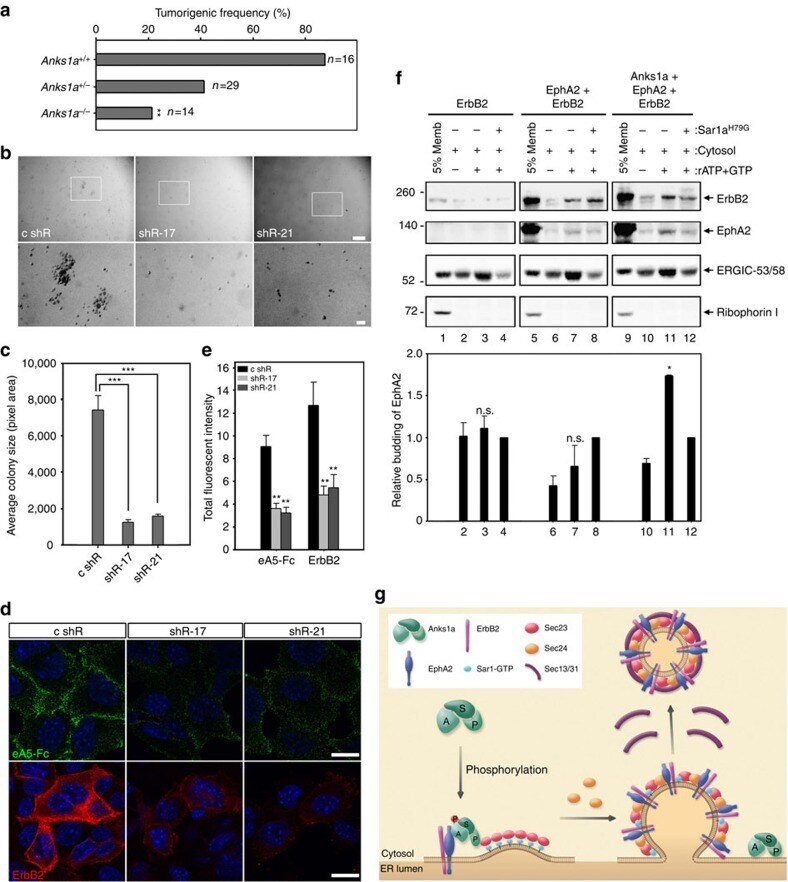
Anks1a regulates COPII-mediated anterograde transport of receptor tyrosine kinases critical for tumorigenesis.
Characterization of a novel angiogenic model based on stable, fluorescently labelled endothelial cell lines amenable to scale-up for high content screening.
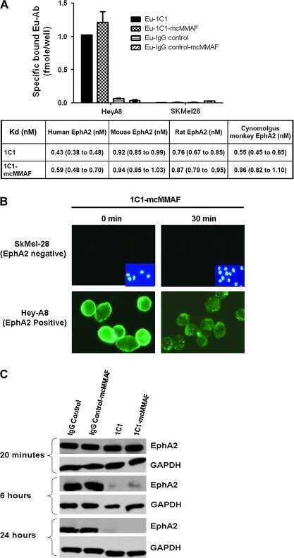
EphA2 immunoconjugate as molecularly targeted chemotherapy for ovarian carcinoma.
A human antibody-drug conjugate targeting EphA2 inhibits tumor growth in vivo.
Framework shuffling of antibodies to reduce immunogenicity and manipulate functional and biophysical properties.
Kaminskyy VO, Hååg P, Novak M, Végvári Á, Arapi V, Lewensohn R, Viktorsson K
Cancers 2021 Feb 28;13(5)
Cancers 2021 Feb 28;13(5)
microRNA-451a promoter methylation regulated by DNMT3B expedites bladder cancer development via the EPHA2/PI3K/AKT axis.
Liu B, Sun W, Gao W, Li L, Cao Z, Yang X, Liu J, Guo Y
BMC cancer 2020 Oct 21;20(1):1019
BMC cancer 2020 Oct 21;20(1):1019
A novel immortalized hepatocyte-like cell line (imHC) supports in vitro liver stage development of the human malarial parasite Plasmodium vivax.
Pewkliang Y, Rungin S, Lerdpanyangam K, Duangmanee A, Kanjanasirirat P, Suthivanich P, Sa-Ngiamsuntorn K, Borwornpinyo S, Sattabongkot J, Patrapuvich R, Hongeng S
Malaria journal 2018 Jan 25;17(1):50
Malaria journal 2018 Jan 25;17(1):50
Ephrin B3 interacts with multiple EphA receptors and drives migration and invasion in non-small cell lung cancer.
Efazat G, Novak M, Kaminskyy VO, De Petris L, Kanter L, Juntti T, Bergman P, Zhivotovsky B, Lewensohn R, Hååg P, Viktorsson K
Oncotarget 2016 Sep 13;7(37):60332-60347
Oncotarget 2016 Sep 13;7(37):60332-60347
Anks1a regulates COPII-mediated anterograde transport of receptor tyrosine kinases critical for tumorigenesis.
Lee H, Noh H, Mun J, Gu C, Sever S, Park S
Nature communications 2016 Sep 13;7:12799
Nature communications 2016 Sep 13;7:12799
Characterization of a novel angiogenic model based on stable, fluorescently labelled endothelial cell lines amenable to scale-up for high content screening.
Prigozhina NL, Heisel A, Wei K, Noberini R, Hunter EA, Calzolari D, Seldeen JR, Pasquale EB, Ruiz-Lozano P, Mercola M, Price JH
Biology of the cell 2011 Oct 1;103(10):467-81
Biology of the cell 2011 Oct 1;103(10):467-81
EphA2 immunoconjugate as molecularly targeted chemotherapy for ovarian carcinoma.
Lee JW, Han HD, Shahzad MM, Kim SW, Mangala LS, Nick AM, Lu C, Langley RR, Schmandt R, Kim HS, Mao S, Gooya J, Fazenbaker C, Jackson D, Tice DA, Landen CN, Coleman RL, Sood AK
Journal of the National Cancer Institute 2009 Sep 2;101(17):1193-205
Journal of the National Cancer Institute 2009 Sep 2;101(17):1193-205
A human antibody-drug conjugate targeting EphA2 inhibits tumor growth in vivo.
Jackson D, Gooya J, Mao S, Kinneer K, Xu L, Camara M, Fazenbaker C, Fleming R, Swamynathan S, Meyer D, Senter PD, Gao C, Wu H, Kinch M, Coats S, Kiener PA, Tice DA
Cancer research 2008 Nov 15;68(22):9367-74
Cancer research 2008 Nov 15;68(22):9367-74
Framework shuffling of antibodies to reduce immunogenicity and manipulate functional and biophysical properties.
Damschroder MM, Widjaja L, Gill PS, Krasnoperov V, Jiang W, Dall'Acqua WF, Wu H
Molecular immunology 2007 Apr;44(11):3049-60
Molecular immunology 2007 Apr;44(11):3049-60
No comments: Submit comment
Supportive validation
- Submitted by
- Invitrogen Antibodies (provider)
- Main image

- Experimental details
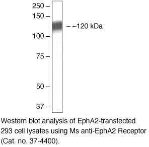
- Western blot analysis of EphA2-transfected 293 cell lysates using Ms anti-EphA2 Receptor (Product # 37-4400).
- Submitted by
- Invitrogen Antibodies (provider)
- Main image

- Experimental details
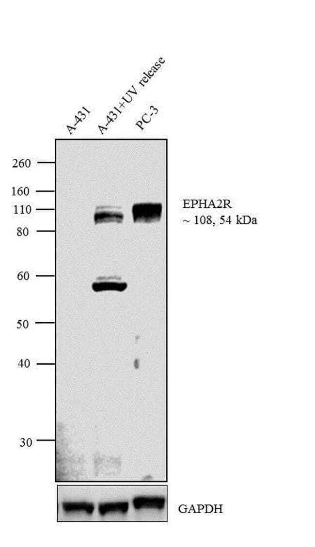
- Western blot analysis was performed on membrane enriched extracts (30 µg lysate) of A-431 (Lane 1), A-431+UV release (5 hour treatment with UVR, after treatment release for 12 hours) (Lane 2) and PC-3 (Lane 3). The blots were probed with Anti-EphA2 Receptor Mouse Monoclonal Antibody (Product # 37-4400, 1-3 µg/mL) and detected by chemiluminescence using Goat anti-Mouse IgG (H+L) Superclonal™ Secondary Antibody, HRP conjugate (Product # A28177, 0.4 µg/mL, 1:2500 dilution). Two isoforms of 108 kDa and 54 kDa corresponding to EphA2 was observed in A431 upon UV treatment and release and one isoform of 108 kDa was observed in PC-3. Known quantity of protein samples were electrophoresed using Novex® NuPAGE® 10 % Bis-Tris gel (Product # NP0301BOX), XCell SureLock™ Electrophoresis System (Product # EI0002) and Novex® Sharp Pre-Stained Protein Standard (Product # LC5800). Resolved proteins were then transferred onto a nitrocellulose membrane with PierceTM Power Blotter System (Product # 22834). The membrane was probed with the relevant primary and secondary Antibody using iBind™ Flex Western Starter Kit (Product # SLF2000S). Chemiluminescent detection was performed using Pierce™ ECL Western Blotting Substrate (Product # 32106).
Supportive validation
- Submitted by
- Invitrogen Antibodies (provider)
- Main image
- Experimental details
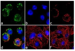
- Immunofluorescent analysis of EphA2 was performed using 70% confluent log phase A549 cells treated with UV-302 nm for 60 minutes. The cells were fixed with 4% paraformaldehyde for 10 minutes, permeabilized with 0.1% Triton™ X-100 for 10 minutes, and blocked with 1% BSA for 1 hour at room temperature. The cells were labeled with EphA2 (1C11A12) Mouse Monoclonal Antibody (Product # 37-4400) at 2 µg/mL in 0.1% BSA and incubated for 3 hours at room temperature and then labeled with Goat anti-Mouse IgG (H+L) Superclonal™ Secondary Antibody, Alexa Fluor® 488 conjugate (Product # A28175) a dilution of 1:2000 for 45 minutes at room temperature (Panel a: green). Nuclei (Panel b: blue) were stained with SlowFade® Gold Antifade Mountant with DAPI (Product # S36938). F-actin (Panel c: red) was stained with Alexa Fluor® 555 Rhodamine Phalloidin (Product # R415, 1:300). Panel d represents the merged image showing cytoplasmic localization. Panel e is untreated cell with no signal. Panel f represents control cells with no primary antibody to assess background. The images were captured at 60X magnification.
Supportive validation
- Submitted by
- Invitrogen Antibodies (provider)
- Main image
- Experimental details
- NULL

- Submitted by
- Invitrogen Antibodies (provider)
- Main image

- Experimental details
- NULL
- Submitted by
- Invitrogen Antibodies (provider)
- Main image

- Experimental details
- Figure 5 Anks1a influences breast tumorigenesis by regulating the ER export of EphA2-ErbB2 complexes. ( a ) Anks1a +/- female mice were crossed with Anks1a +/- ; MMTV-Neu male mice to obtain female mice with the indicated genotypes. All experiments monitoring breast tumour formation were carried out in mice of the FVB genetic background ( n =16, 29, 14 per genotype). ** P
- Submitted by
- Invitrogen Antibodies (provider)
- Main image
- Experimental details
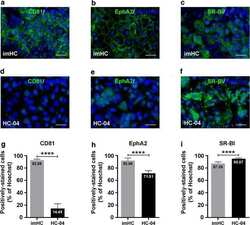
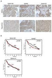
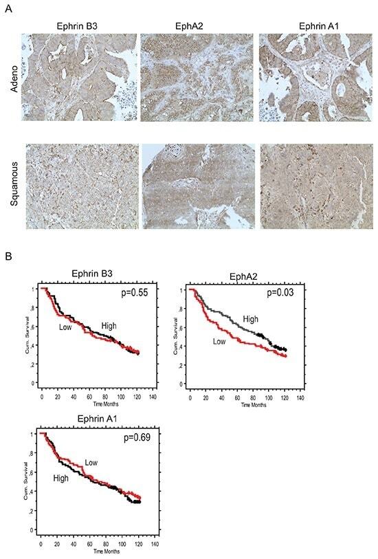
- Figure 5 Concomitant Ephrin B3, Ephrin A1 and EphA2 expression in NSCLC clinical specimens Ephrin B3, Ephrin A1 and EphA2 expression levels and their relation to patient overall survival (OS) was analyzed in a cohort of Stage IA-Stage IB NSCLC specimen using immunohistochemistry. A. Examples of Ephrin B3, EphA2 and Ephrin A1 staining in adenocarcinoma and squamous cell carcinoma NSCLC specimens. 20x magnification. B. Kaplan-Maier curves showing the association between Ephrin B3, EphA2 and Ephrin A1 expression intensity levels and OS in the NSCLC patient cohort. Red line: Low score, black line: High score. Wilcoxon test was used for statistical assessment and the p-values are indicated.
- Submitted by
- Invitrogen Antibodies (provider)
- Main image
- Experimental details
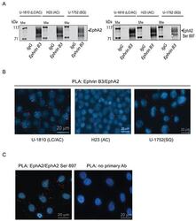
- Figure 3 Ephrin B3 is a ligand of multiple EphA receptors The interactions of Ephrin B3 with Ephs were studied by immunoprecipitation in untreated NSCLC cells as indicated. A. Ephrin B3was immunoprecipitated from lysates of U-1810, H23, and U-1752 cells and the resulting immunocomplexes studied in western blot by antibodies against total EphA2 or EphA2 Ser897. B. The interaction between Ephrin B3 and EphA2 were analyzed by PLA in situ in untreated U-1810, H23 or U1-752 cells. Slides were incubated with Ephrin B3 and EphA2 antibodies and probed with Texas red (red) PLA probes with DAPI (blue) used for counterstaining cell nuclei. C. The interaction between total EphA2 and Ser897 phosphorylated EphA2 was verified with PLA in U-1810 cells. Texas-red labelled PLA probes were added (red) with DAPI (blue) used for counterstaining of nuclei. As a control U-1810 cells were used in PLA reactions omitting the primary antibodies. D. NSCLC cell lines were profiled for the expression of EphA2 Ser897, using western blotting. To control equal loading among the samples, beta-Tubulin was used. E. The interactions of Ephrin B3 with Ephs were studied by immunoprecipitation in untreated NSCLC cells as indicated. Ephrin B3was immunoprecipitated from cell lysates of U-1810, H23, U-1810 and U-1752 and the resulting immunocomplexes analyzed by western blot with antibodies against EphA3, EphA4 and EphA5.

- Submitted by
- Invitrogen Antibodies (provider)
- Main image
- Experimental details
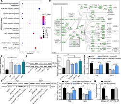
- Fig. 4 miR-451a targets EPHA2 to regulate the PI3K/AKT signaling. a , the targeting mRNAs of miR-451a predicted by StarBase and analyzed using a DAVID 6.8 online bioinformatics analysis tool. Bubble plots showed 14 signaling pathways; b , gene distribution in the PI3K/AKT signaling in the KEGG database; c , EPHA2 mRNA expression in BCa cell lines T24, 5637, TCCSUP, UM-UC3 and SV-HUC-1 cells determined by RT-qPCR; d , EPHA2 protein expression in BCa cell lines T24, 5637, TCCSUP, UM-UC3 and SV-HUC-1 cells determined by western blot (full-length blots/gels are presented in Supplementary Figure 2 A, B); e , EPHA2 mRNA expression in BCa cell lines after transfection determined by RT-qPCR; f , EPHA2 protein expression in BCa cell lines after transfection determined by western blot (full-length blots/gels are presented in Supplementary Figure 3 A-D); g , the binding relationship between miR-451a and EPHA2 verified by dual-luciferase assays. Statistically significant differences were calculated using one-way or two-way ANOVA, followed by Tukey's multiple comparison test. ** p < 0.01 vs sh-NC or sh-DNMT3B + inhibitor NC treatment, *** p < 0.001 vs SV-HUC-1 cells
