Explore
 Validate
 Learn
Learn Western blot
 Immunocytochemistry
Antibody data
- Antibody Data
- Antigen structure
- References [0]
- Comments [0]
- Validations
- Western blot [1]
- Immunohistochemistry [1]
- Flow cytometry [1]
Submit
Validation data
Reference
Comment
Report error
- Product number
- MAB91671-100 - Provider product page

- Provider
- Novus Biologicals
- Product name
- Mouse Monoclonal Neutrophil Elastase/ELA2 Antibody
- Antibody type
- Monoclonal
- Description
- Protein A or G purified from hybridoma culture supernatant. Detects human ELA2 in direct ELISAs and Western blots.
- Reactivity
- Human
- Host
- Mouse
- Conjugate
- Unconjugated
- Isotype
- IgG
- Vial size
- 100 ug
- Storage
- Use a manual defrost freezer and avoid repeated freeze-thaw cycles. 12 months from date of receipt, -20 to -70 degreesC as supplied. 1 month, 2 to 8 degreesC under sterile conditions after reconstitution. 6 months, -20 to -70 degreesC under sterile conditions after reconstitution.
No comments: Submit comment
Supportive validation
- Submitted by
- Novus Biologicals (provider)
- Main image
- Experimental details
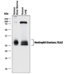
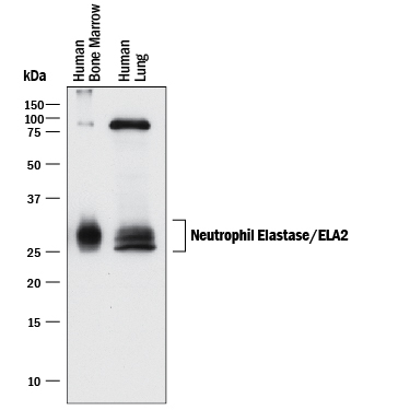
- Detection of Human Neutrophil Elastase/ELA2 by Western Blot. Western blot shows lysates of human bone marrow and human lung tissue. PVDF membrane was probed with 0.1 µg/mL of Mouse Anti-Human Neutrophil Elastase/ELA2 Monoclonal Antibody (Catalog # MAB91671) followed by HRP-conjugated Anti-Mouse IgG Secondary Antibody (Catalog # HAF018). Specific bands were detected for Neutrophil Elastase/ELA2 at approximately 25-30 kDa (as indicated). This experiment was conducted under reducing conditions and using Immunoblot Buffer Group 1.
Supportive validation
- Submitted by
- Novus Biologicals (provider)
- Main image
- Experimental details
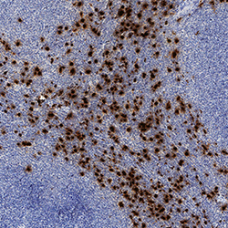
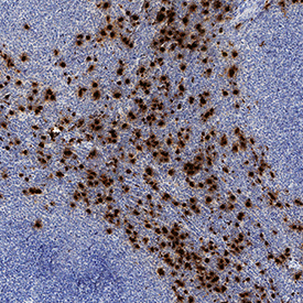
- Neutrophil Elastase/ELA2 in Human Lymphoma. Neutrophil Elastase/ELA2 was detected in immersion fixed paraffin-embedded sections of human lymphoma using Mouse Anti-Human Neutrophil Elastase/ELA2 Monoclonal Antibody (Catalog # MAB91671) at 5 µg/mL for 1 hour at room temperature followed by incubation with the Anti-Mouse IgG VisUCyte™ HRP Polymer Antibody (Catalog # VC001). Tissue was stained using DAB (brown) and counterstained with hematoxylin (blue). Specific staining was localized to cytoplasm. View our protocol for IHC Staining with VisUCyte HRP Polymer Detection Reagents.
Supportive validation
- Submitted by
- Novus Biologicals (provider)
- Main image
- Experimental details
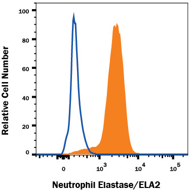
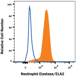
- Detection of Neutrophil Elastase/ELA2 in THP-1 Human Cell Line by Flow Cytometry. THP-1 human acute monocytic leukemia cell line treated with 3 μM monensin for 3 hours was stained with Mouse Anti-Human Neutrophil Elastase/ELA2 Monoclonal Antibody (Catalog # MAB91671, filled histogram) or isotype control antibody (Catalog # MAB002, open histogram), followed by Phycoerythrin-conjugated Anti-Mouse IgG Secondary Antibody (Catalog # F0102B). To facilitate intracellular staining, cells were fixed with Flow Cytometry Fixation Buffer (Catalog # FC004) and permeabilized with Flow Cytometry Permeabilization/Wash Buffer I (Catalog # FC005). View our protocol for Staining Intracellular Molecules.