 Explore
 Validate
 Learn
Learn Western blot
Antibody data
- Antibody Data
- Antigen structure
- References [0]
- Comments [0]
- Validations
- Western blot [3]
- Immunocytochemistry [1]
Submit
Validation data
Reference
Comment
Report error
- Product number
- PA5-21457 - Provider product page

- Provider
- Invitrogen Antibodies
- Product name
- RAP1A Polyclonal Antibody
- Antibody type
- Polyclonal
- Antigen
- Recombinant protein fragment
- Description
- Recommended positive controls: 293T, A431, H1299, HeLa, HepG2, Molt-4, Raji.
- Concentration
- 0.53 mg/mL
No comments: Submit comment
Supportive validation
- Submitted by
- Invitrogen Antibodies (provider)
- Main image
- Experimental details
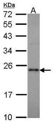
- Western blot analysis of RAP1A using 30 µg of MOLT4 lysate. Samples were loaded onto a 12% SDS-PAGE gel and probed with a RAP1A polyclonal antibody (Product # PA5-21457) at a dilution of 1:1000.

- Submitted by
- Invitrogen Antibodies (provider)
- Main image

- Experimental details
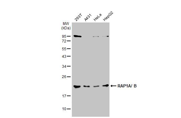
- Western Blot using RAP1A Polyclonal Antibody (Product # PA5-21457). Various whole cell extracts (30 µg) were separated by 12% SDS-PAGE, and the membrane was blotted with RAP1A Polyclonal Antibody (Product # PA5-21457) diluted at 1:1,000. The HRP-conjugated anti-rabbit IgG antibody was used to detect the primary antibody.
- Submitted by
- Invitrogen Antibodies (provider)
- Main image

- Experimental details
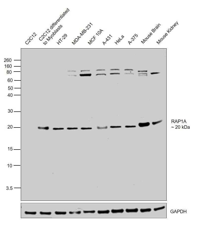
- Western blot was performed using Anti-RAP1A Polyclonal Antibody (Product # PA5-21457) and 20 kDa band corresponding to RAP1A was observed across the cell lines and tissues tested except C2C12 in which the expression was observed upon myoblast differentiation. Uncharacterized bands of ~75 and 80 kDa was also observed. Whole cell lysates (30ug lysate) of C2C12 (Lane 1), C2C12 cells differentiated to Myoblasts (Lane2), HT-29 (Lane 3), MDA-MB-231 (Lane 4), MCF 10A (Lane 5), A-431 (Lane 6), HeLa (Lane 7) and A-375 (Lane 8), tissue extracts (30 µg lysate) of Mouse Brain (Lane 9) and Mouse Kidney (Lane 10) were electrophoresed using Novex® NuPAGE® 4-12 % Bis-Tris gel (Product # NP0322BOX). Resolved proteins were then transferred onto a nitrocellulose membrane (Product # IB23001) by iBlot® 2 Dry Blotting System (Product # IB21001). The blot was probed with the primary antibody (1:1000 dilution) and detected by chemiluminescence with Goat anti-Rabbit IgG (H+L), Superclonal™ Recombinant Secondary Antibody, HRP (Product # A27036, 1:4000 dilution) using the iBright FL 1000 (Product # A32752). Chemiluminescent detection was performed using Novex® ECL Chemiluminescent Substrate Reagent Kit (Product # WP20005).
Supportive validation
- Submitted by
- Invitrogen Antibodies (provider)
- Main image
- Experimental details
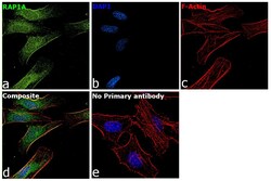
- Immunofluorescence analysis of RAP1A was performed using 70% confluent log phase HeLa cells. The cells were fixed with 4% paraformaldehyde for 10 minutes, permeabilized with 0.1% Triton™ X-100 for 15 minutes, and blocked with 2% BSA for 1 hour at room temperature. The cells were labeled with RAP1A Polyclonal Antibody (Product PA5-21457) at 1 µg/mL in 0.1% BSA, incubated at 4 degree Celsius overnight and then with Goat anti-Rabbit IgG (H+L), Superclonal™ Recombinant Secondary Antibody, Alexa Fluor 488 (Product # A27034, 1:2000 dilution) for 45 minutes at room temperature (Panel a: Green). Nuclei (Panel b: Blue) were stained with SlowFade® Gold Antifade Mountant with DAPI (Product # S36938). F-actin (Panel c: Red) was stained with Rhodamine Phalloidin (Product # R415, 1:300). Panel d represents the merged image showing membrane and cytoplasmic localization. Panel e represents control cells with no primary antibody to assess background. The images were captured at 60X magnification.
