Explore
 Validate
 Learn
Learn Western blot
Antibody data
- Antibody Data
- Antigen structure
- References [0]
- Comments [0]
- Validations
- Western blot [2]
- Immunocytochemistry [2]
- Immunohistochemistry [1]
- Flow cytometry [2]
Submit
Validation data
Reference
Comment
Report error
- Product number
- PA5-17937 - Provider product page

- Provider
- Invitrogen Antibodies
- Product name
- GADD34 Polyclonal Antibody
- Antibody type
- Polyclonal
- Antigen
- Synthetic peptide
- Reactivity
- Human
- Host
- Goat
- Isotype
- IgG
- Vial size
- 100 µg
- Concentration
- 0.5 mg/mL
- Storage
- -20° C, Avoid Freeze/Thaw Cycles
No comments: Submit comment
Supportive validation
- Submitted by
- Invitrogen Antibodies (provider)
- Main image
- Experimental details
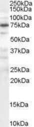
- Western Blot staining of HepG2 cell lysate using Product # PA5-17937 at a concentration of 0.1 µg/mL, the primary antibody incubation was 1 hour and the detection method was chemiluminescence.
- Submitted by
- Invitrogen Antibodies (provider)
- Main image
- Experimental details
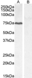
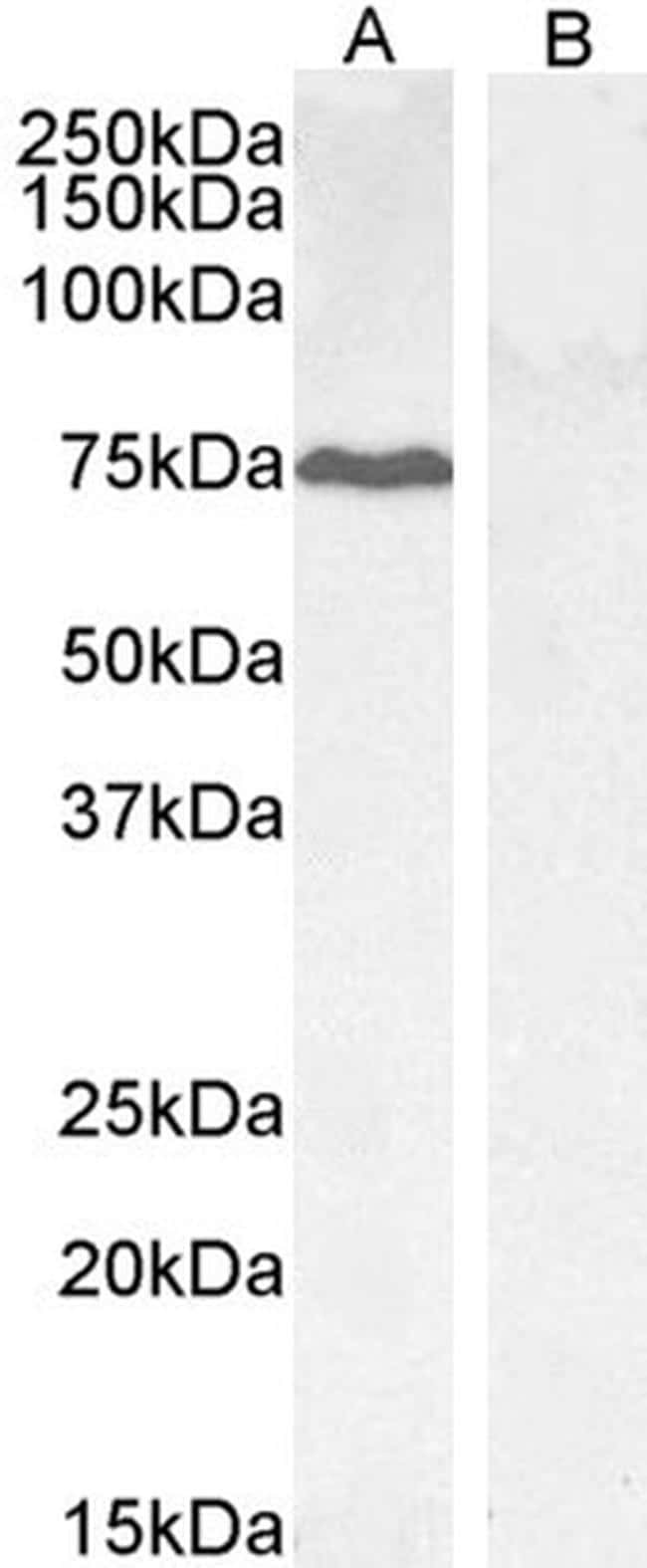
- Western blot analysis of GADD34 by a GADD34 monoclonal antibody (Product # PA5-17937) at a concentration of 0.3 µg/mL. HepG2 (A) and negative control KLY (B) lysate (35µg protein in RIPA buffer) Detected by chemiluminescence.
Supportive validation
- Submitted by
- Invitrogen Antibodies (provider)
- Main image
- Experimental details
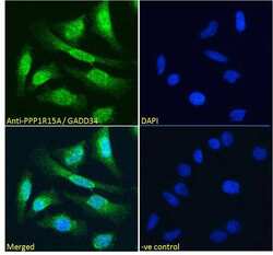
- Immunofluorescence analysis of GADD34 in HeLa cells using a GADD34 monoclonal antibody (Product # PA5-17937) at 10 µg/mL for1hr. The cells were paraformaldehyde fixed and permeabilized with 0.15% Triton. Primary incubation was followed by Alexa Fluor 488 secondary antibody (4 µg/mL) showing strong nuclear and some cytoplasmic staining. The nuclear stain is DAPI (blue). Negative control: Unimmunized goat IgG (10 µg/mL)followed by Alexa Fluor 488 secondary antibody (4 µg/mL).

- Submitted by
- Invitrogen Antibodies (provider)
- Main image

- Experimental details
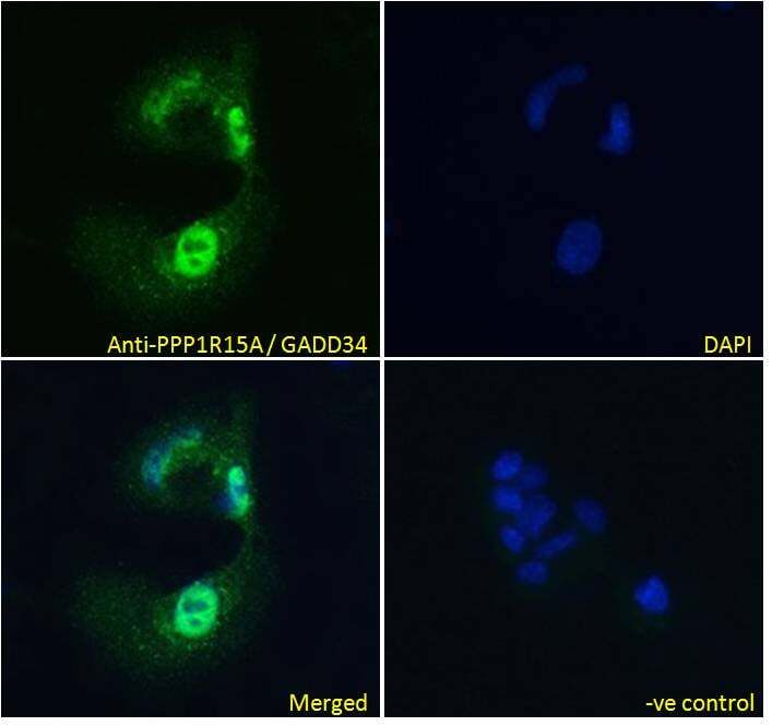
- Immunofluorescence analysis of GADD34 in HepG2 cells using a GADD34 monoclonal antibody (Product # PA5-17937) at 10 µg/mL for1hr. The cells were paraformaldehyde fixed and permeabilized with 0.15% Triton. Primary incubation was followed by Alexa Fluor 488 secondary antibody (4 µg/mL) showing strong nuclear and some cytoplasmic staining. The nuclear stain is DAPI (blue). Negative control: Unimmunized goat IgG (10 µg/mL)followed by Alexa Fluor 488 secondary antibody (4 µg/mL).
Supportive validation
- Submitted by
- Invitrogen Antibodies (provider)
- Main image

- Experimental details
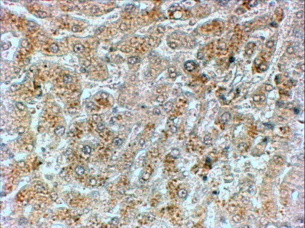
- Immunohistochemical analysis of GADD34 in paraffin embedded human liver using a GADD34 polyclonal antibody (Product #PA5-17937) at a concentration of 2 µg/mL. Steamed antigen retrieval was performed with pH 6 buffered citrate. Samples were then stained with horseradish peroxidase.
Supportive validation
- Submitted by
- Invitrogen Antibodies (provider)
- Main image
- Experimental details
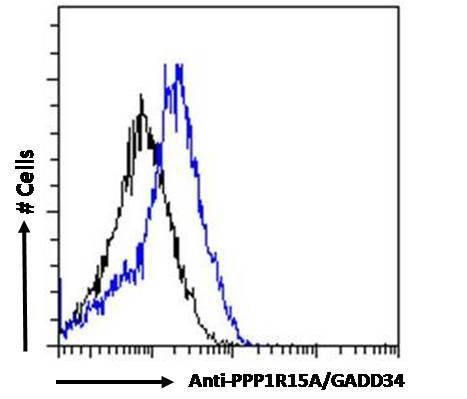
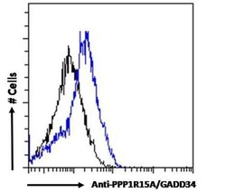
- Flow cytometric analysis of GADD34 in HepG2 cells using a GADD34 monoclonal antibody (Product # PA5-17937) at 10 µg/mL for 1hr, depicted by the blue line. The cells were paraformaldehyde fixed and permeabilized with 0.5% Triton. Primary incubation followed by Alexa Fluor 488 secondary antibody (0.4 µg/mL). IgG control: Unimmunized goat IgG (black line) followed by Alexa Fluor 488 secondary antibody.

- Submitted by
- Invitrogen Antibodies (provider)
- Main image
- Experimental details
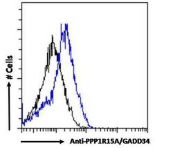
- Flow cytometric analysis of GADD34 in HepG2 cells using a polyclonal antibody (Product #PA5-17937). HepG2 cells (blue line) were paraformaldehyde fixed and permeabilized with 0.5% Triton. The primary antibody was incubated for one hour (10 µg/mL) followed by an Alexa Fluor 488 secondary antibody (0.4 µg/mL). IgG control: Unimmunized goat IgG (black line) followed by an Alexa Fluor 488 secondary antibody.