 Explore
 Validate
 Learn
Learn Western blot
Antibody data
- Antibody Data
- Antigen structure
- References [0]
- Comments [0]
- Validations
- Western blot [4]
- Flow cytometry [1]
Submit
Validation data
Reference
Comment
Report error
- Product number
- 44-688G - Provider product page

- Provider
- Invitrogen Antibodies
- Product name
- ERK5 Polyclonal Antibody
- Antibody type
- Polyclonal
- Antigen
- Synthetic peptide
- Reactivity
- Human, Mouse, Rat
- Host
- Rabbit
- Isotype
- IgG
- Vial size
- 100 µL
- Storage
- -20°C
No comments: Submit comment
Supportive validation
- Submitted by
- Invitrogen Antibodies (provider)
- Main image

- Experimental details
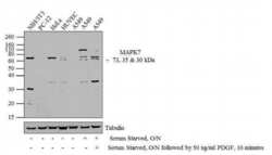
- Western blot analysis was performed on whole cell extracts (20 µg lysate) of NIH/3T3 (Lane 1), PC-12 (Lane 2), HeLa (lane 3), HUVEC (lane 4), A549 (lane 5), Serum Starved A549 (lane 6) and A549 serum starved overnight followed by treatment for 10 minutes with 50 ng/mL of PDGF (lane 7). The blots were probed with Anti-MAPK7 Rabbit Polyclonal Antibody (Product # 44-688G, 1:500-1:2000 dilution) and detected by chemiluminescence using Goat anti-Rabbit IgG (H+L) Superclonal™ Secondary Antibody, HRP conjugate (Product # A27036, 0.4 µg/mL, 1:2500 dilution). Three bands ~ 73, 35 and 30 kDa corresponding to MAPK7 was observed across the cell lines tested, additional non-specific band aroudn 110 kDa is seen in A549 serum starved cells . Known quantity of protein samples were electrophoresed using Novex® NuPAGE® 12 % Bis-Tris gel (Product # NP0342BOX), XCell SureLock™ Electrophoresis System (Product # EI0002) and Novex® Sharp Pre-Stained Protein Standard (Product # LC5800). Resolved proteins were then transferred onto a nitrocellulose membrane with iBlot® 2 Dry Blotting System (Product # IB21001). The membrane was probed with the relevant primary and secondary Antibody following blocking with 5 % skimmed milk. Chemiluminescent detection was performed using Pierce™ ECL Western Blotting Substrate (Product # 32106).

- Submitted by
- Invitrogen Antibodies (provider)
- Main image
- Experimental details
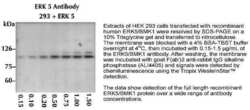
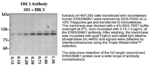
- Western blot analysis of ERK5 using a polyclonal antibody (Product # 44-688G).
- Submitted by
- Invitrogen Antibodies (provider)
- Main image
- Experimental details
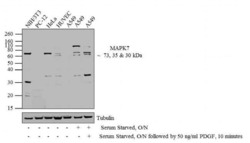
- Western blot analysis was performed on whole cell extracts (20 µg lysate) of NIH/3T3 (Lane 1), PC-12 (Lane 2), HeLa (lane 3), HUVEC (lane 4), A549 (lane 5), Serum Starved A549 (lane 6) and A549 serum starved overnight followed by treatment for 10 minutes with 50 ng/mL of PDGF (lane 7). The blots were probed with Anti-MAPK7 Rabbit Polyclonal Antibody (Product # 44-688G, 1:500-1:2000 dilution) and detected by chemiluminescence using Goat anti-Rabbit IgG (H+L) Superclonal™ Secondary Antibody, HRP conjugate (Product # A27036, 0.4 µg/mL, 1:2500 dilution). Three bands ~ 73, 35 and 30 kDa corresponding to MAPK7 was observed across the cell lines tested, additional non-specific band aroudn 110 kDa is seen in A549 serum starved cells . Known quantity of protein samples were electrophoresed using Novex® NuPAGE® 12 % Bis-Tris gel (Product # NP0342BOX), XCell SureLock™ Electrophoresis System (Product # EI0002) and Novex® Sharp Pre-Stained Protein Standard (Product # LC5800). Resolved proteins were then transferred onto a nitrocellulose membrane with iBlot® 2 Dry Blotting System (Product # IB21001). The membrane was probed with the relevant primary and secondary Antibody following blocking with 5 % skimmed milk. Chemiluminescent detection was performed using Pierce™ ECL Western Blotting Substrate (Product # 32106).

- Submitted by
- Invitrogen Antibodies (provider)
- Main image
- Experimental details
- Western blot analysis was performed on whole cell extracts (20 µg lysate) of NIH/3T3 (Lane 1), PC-12 (Lane 2), HeLa (lane 3), HUVEC (lane 4), A549 (lane 5), Serum Starved A549 (lane 6) and A549 serum starved overnight followed by treatment for 10 minutes with 50 ng/mL of PDGF (lane 7). The blots were probed with Anti-MAPK7 Rabbit Polyclonal Antibody (Product # 44-688G, 1:500-1:2000 dilution) and detected by chemiluminescence using Goat anti-Rabbit IgG (H+L) Superclonal™ Secondary Antibody, HRP conjugate (Product # A27036, 0.4 µg/mL, 1:2500 dilution). Three bands ~ 73, 35 and 30 kDa corresponding to MAPK7 was observed across the cell lines tested, additional non-specific band aroudn 110 kDa is seen in A549 serum starved cells . Known quantity of protein samples were electrophoresed using Novex® NuPAGE® 12 % Bis-Tris gel (Product # NP0342BOX), XCell SureLock™ Electrophoresis System (Product # EI0002) and Novex® Sharp Pre-Stained Protein Standard (Product # LC5800). Resolved proteins were then transferred onto a nitrocellulose membrane with iBlot® 2 Dry Blotting System (Product # IB21001). The membrane was probed with the relevant primary and secondary Antibody following blocking with 5 % skimmed milk. Chemiluminescent detection was performed using Pierce™ ECL Western Blotting Substrate (Product # 32106).

Supportive validation
- Submitted by
- Invitrogen Antibodies (provider)
- Main image
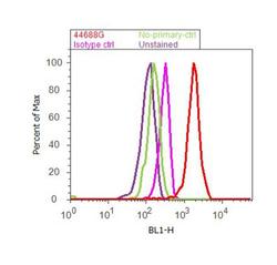
- Experimental details
- Flow cytometry analysis of MAPK7 was done on NIH/3T3 cells. Cells were fixed with 70% ethanol for 10 minutes, permeabilized with 0.25% Triton™ X-100 for 20 minutes, and blocked with 5% BSA for 30 minutes at room temperature. Cells were labeled with MAPK7 Rabbit Polyclonal Antibody (44688G, red histogram) or with rabbit isotype control (pink histogram) at 3-5 ug/million cells in 2.5% BSA. After incubation at room temperature for 2 hours, the cells were labeled with Alexa Fluor® 488 Goat Anti-Rabbit Secondary Antibody (A11008) at a dilution of 1:400 for 30 minutes at room temperature. The representative 10,000 cells were acquired and analyzed for each sample using an Attune® Acoustic Focusing Cytometer. The purple histogram represents unstained control cells and the green histogram represents no-primary-antibody control.
