Explore
 Validate
 Learn
LearnMA5-12333
antibody from Invitrogen Antibodies
Targeting: MCM2
BM28, CCNL1, cdc19, CDCL1, D3S3194, DFNA70, KIAA0030
 Western blot
Antibody data
- Antibody Data
- Antigen structure
- References [1]
- Comments [0]
- Validations
- Western blot [4]
- Immunohistochemistry [1]
Submit
Validation data
Reference
Comment
Report error
- Product number
- MA5-12333 - Provider product page

- Provider
- Invitrogen Antibodies
- Product name
- MCM2 Monoclonal Antibody (CRCT2.1)
- Antibody type
- Monoclonal
- Antigen
- Other
- Description
- MA5-12333 targets MCM2 in IHC (P) and WB applications and shows reactivity with Human samples.
- Antibody clone number
- CRCT2.1
- Concentration
- 0.4 mg/mL
Submitted references Differential biomarker expression in head and neck cancer correlates with anatomical localization.
Tamás L, Szentkúti G, Eros M, Dános K, Brauswetter D, Szende B, Zsákovics I, Krenács T
Pathology oncology research : POR 2011 Sep;17(3):721-7
Pathology oncology research : POR 2011 Sep;17(3):721-7
No comments: Submit comment
Supportive validation
- Submitted by
- Invitrogen Antibodies (provider)
- Main image
- Experimental details
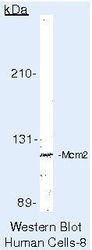
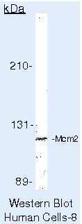
- Western blot of MCM2 using MCM2 Monoclonal Antibody (Product # MA5-12333) on HeLa Cells.
- Submitted by
- Invitrogen Antibodies (provider)
- Main image
- Experimental details
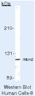
- Western blot of MCM2 using MCM2 Monoclonal Antibody (Product # MA5-12333) on HeLa Cells.
- Submitted by
- Invitrogen Antibodies (provider)
- Main image
- Experimental details
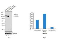
- Knockdown of MCM2 was achieved by transfecting MCF7 cells with MCM2 specific siRNA (Silencer® select Product # s8586 and s8587). Western blot analysis (Fig a) was performed using modified whole cell extract (1% SDS) from the MCM2 knock down cells (lane 3), non-specific scrambled siRNA transfected cells (lane 2) and untransfected cells (lane 1). The blots were probed with Anti-MCM2 Mouse monoclonal antibody (Product # MA5-12333, 2 µg/mL) and Goat anti-Mouse IgG (H+L) Secondary Antibody, HRP conjugate (Product # 62-6520, 1:4000 dilution). Densitometric analysis of this western blot is shown in histogram (Fig b). Loss of signal upon siRNA mediated knock down confirms that antibody is specific to MCM2.

- Submitted by
- Invitrogen Antibodies (provider)
- Main image
- Experimental details
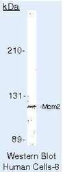
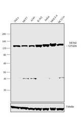
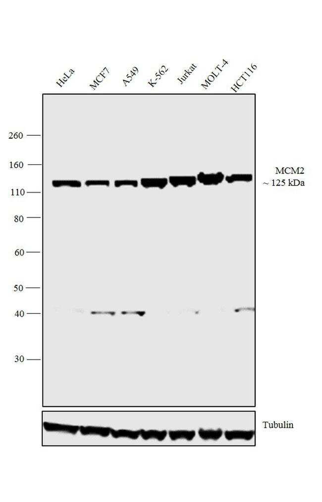
- Western blot analysis was performed on nuclear enriched extracts (30 µg lysate) of HeLa (Lane 1), MCF7 (Lane 2), A549 (Lane3), K-562 (Lane 4), Jurkat (Lane 5), MOLT4 (Lane 6) and HCT116 (Lane 7). The blot was probed with Mouse anti-MCM2 Monoclonal Antibody (Product # MA5-12333, 1 µg/mL) and detected by chemiluminescence using Goat anti-Mouse IgG (H+L) Superclonal™ Secondary Antibody, HRP conjugate (Product # A28177, 0.25 µg/mL, 1:4000 dilution). A 125 kDa band corresponding to MCM2 was observed across the cell lines tested. Known quantity of protein samples were electrophoresed using Novex® NuPAGE® 4-12% Bis-Tris gel (Product # NP0321BOX), XCell SureLock™ Electrophoresis System (Product # EI0002) and Novex® Sharp Pre-Stained Protein Standard (Product # LC5800). Resolved proteins were then transferred onto a nitrocellulose membrane with iBlot® 2 Dry Blotting System (Product # IB21001). The membrane was probed with the relevant primary and secondary Antibody following blocking with 5 % skimmed milk. Chemiluminescent detection was performed using Pierce™ ECL Western Blotting Substrate (Product # 32106).
Supportive validation
- Submitted by
- Invitrogen Antibodies (provider)
- Main image

- Experimental details
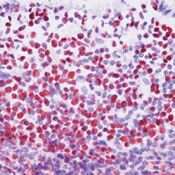
- Formalin-fixed, paraffin-embedded human tonsil stained with MCM2 using peroxidase-conjugate and AEC chromogen. Note nuclear staining of lymphocytes