Explore
 Validate
 Learn
Learn Flow cytometry
Antibody data
- Antibody Data
- Antigen structure
- References [6]
- Comments [0]
- Validations
- Flow cytometry [1]
Submit
Validation data
Reference
Comment
Report error
- Product number
- 25-9829-41 - Provider product page

- Provider
- Invitrogen Antibodies
- Product name
- Anti-LAP (Latency Associated peptide) Monoclonal Antibody (FNLAP), PE-Cyanine7, eBioscience™
- Antibody type
- Monoclonal
- Antigen
- Other
- Description
- Description: The FNLAP monoclonal antibody reacts with human latency associated peptide (LAP, pro-TGF beta 1, LAP/TGF beta 1). Many different cells produce TGF beta and it mediates effects on the proliferation, differentiation and function of many cell types. TGF beta is synthesized as a precursor that contains LAP at the N-terminus and mature TGF beta at the C-terminus. Processing and cleavage of the precursor protein between amino acids 278 and 279 results in the formation of LAP dimers and TGF beta dimers that then non-covalently associate with each other to form the small latent TGF beta complex. LAP is secreted and can be found in the extracellular matrix. In addition, LAP can also be expressed on platelets and activated regulatory T cells. It is believed that this surface-expressed LAP is due to the binding of LAP to GARP (LRRC32), which is a transmembrane protein that is also found at high levels on platelets and activated regulatory T cells. Applications Reported: This FNLAP antibody has been reported for use in flow cytometric analysis. Applications Tested: This FNLAP antibody has been pre-titrated and tested by flow cytometric analysis of stimulated human peripheral blood cells. This can be used at 5 µL (0.25 µg) per test. A test is defined as the amount (µg) of antibody that will stain a cell sample in a final volume of 100 µL. Cell number should be determined empirically but can range from 10^5 to 10^8 cells/test. Light sensitivity: This tandem dye is sensitive photo-induced oxidation. Please protect this vial and stained samples from light. Fixation: Samples can be stored in IC Fixation Buffer (cat. 00-8222) (100 µL cell sample + 100 µL IC Fixation Buffer) or 1-step Fix/Lyse Solution (cat. 00-5333) for up to 3 days in the dark at 4°C with minimal impact on brightness and FRET efficiency/compensation. Some generalizations regarding fluorophore performance after fixation can be made, but clone specific performance should be determined empirically. Excitation: 488-561 nm; Emission: 775 nm; Laser: Blue Laser, Green Laser, Yellow-Green Laser. Filtration: 0.2 µm post-manufacturing filtered.
- Reactivity
- Human
- Host
- Mouse
- Isotype
- IgG
- Antibody clone number
- FNLAP
- Vial size
- 25 Tests
- Concentration
- 5 µL/Test
- Storage
- 4° C, store in dark, DO NOT FREEZE!
Submitted references Systematic testing and specificity mapping of alloantigen-specific chimeric antigen receptors in regulatory T cells.
Circulating gluten-specific FOXP3+CD39+ regulatory T cells have impaired suppressive function in patients with celiac disease.
NOTCH1 mediates a switch between two distinct secretomes during senescence.
Alloantigen-specific regulatory T cells generated with a chimeric antigen receptor.
Hypomethylation at the regulatory T cell-specific demethylated region in CD25hi T cells is decoupled from FOXP3 expression at the inflamed site in childhood arthritis.
Phenotypic and functional characteristics of CD4+ CD39+ FOXP3+ and CD4+ CD39+ FOXP3neg T-cell subsets in cancer patients.
Dawson NA, Lamarche C, Hoeppli RE, Bergqvist P, Fung VC, McIver E, Huang Q, Gillies J, Speck M, Orban PC, Bush JW, Mojibian M, Levings MK
JCI insight 2019 Mar 21;4(6)
JCI insight 2019 Mar 21;4(6)
Circulating gluten-specific FOXP3+CD39+ regulatory T cells have impaired suppressive function in patients with celiac disease.
Cook L, Munier CML, Seddiki N, van Bockel D, Ontiveros N, Hardy MY, Gillies JK, Levings MK, Reid HH, Petersen J, Rossjohn J, Anderson RP, Zaunders JJ, Tye-Din JA, Kelleher AD
The Journal of allergy and clinical immunology 2017 Dec;140(6):1592-1603.e8
The Journal of allergy and clinical immunology 2017 Dec;140(6):1592-1603.e8
NOTCH1 mediates a switch between two distinct secretomes during senescence.
Hoare M, Ito Y, Kang TW, Weekes MP, Matheson NJ, Patten DA, Shetty S, Parry AJ, Menon S, Salama R, Antrobus R, Tomimatsu K, Howat W, Lehner PJ, Zender L, Narita M
Nature cell biology 2016 Sep;18(9):979-92
Nature cell biology 2016 Sep;18(9):979-92
Alloantigen-specific regulatory T cells generated with a chimeric antigen receptor.
MacDonald KG, Hoeppli RE, Huang Q, Gillies J, Luciani DS, Orban PC, Broady R, Levings MK
The Journal of clinical investigation 2016 Apr 1;126(4):1413-24
The Journal of clinical investigation 2016 Apr 1;126(4):1413-24
Hypomethylation at the regulatory T cell-specific demethylated region in CD25hi T cells is decoupled from FOXP3 expression at the inflamed site in childhood arthritis.
Bending D, Pesenacker AM, Ursu S, Wu Q, Lom H, Thirugnanabalan B, Wedderburn LR
Journal of immunology (Baltimore, Md. : 1950) 2014 Sep 15;193(6):2699-708
Journal of immunology (Baltimore, Md. : 1950) 2014 Sep 15;193(6):2699-708
Phenotypic and functional characteristics of CD4+ CD39+ FOXP3+ and CD4+ CD39+ FOXP3neg T-cell subsets in cancer patients.
Schuler PJ, Schilling B, Harasymczuk M, Hoffmann TK, Johnson J, Lang S, Whiteside TL
European journal of immunology 2012 Jul;42(7):1876-85
European journal of immunology 2012 Jul;42(7):1876-85
No comments: Submit comment
Supportive validation
- Submitted by
- Invitrogen Antibodies (provider)
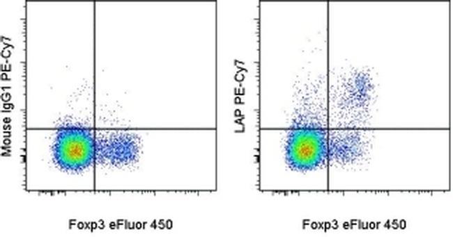
- Main image
- Experimental details
- Normal human peripheral blood cells were stimulated with Anti-Human CD3, Anti-Human CD28 and Human IL-2 recombinant protein for 1 day, and then stained with Anti-Human CD4 FITC (Product # 11-0048-42), and Mouse IgG1 K Isotype Control PE-Cyanine7 (Product # 25-4714-80) (left) or Anti-Human LAP (Latency Associated Peptide) PE-Cyanine7 (right) followed by intracellular staining with Anti-Human Foxp3 eFluor® 450 (Product # 48-4776-42) using Foxp3 Fixation/Permeabilization Buffer and protocol. CD4+ cells in the lymphocyte gate were used for analysis.