Explore
 Validate
 Learn
Learn Immunohistochemistry
Antibody data
- Antibody Data
- Antigen structure
- References [2]
- Comments [0]
- Validations
- Immunohistochemistry [1]
- Other assay [3]
Submit
Validation data
Reference
Comment
Report error
- Product number
- MA5-11748 - Provider product page

- Provider
- Invitrogen Antibodies
- Product name
- Myosin Skeletal Muscle Monoclonal Antibody (MYSN02 (MY-32))
- Antibody type
- Monoclonal
- Antigen
- Other
- Description
- MA5-11748 targets Myosin Skeletal Muscle in IHC (P) applications and shows reactivity with Human samples.
- Antibody clone number
- MYSN02 (MY-32)
- Concentration
- Conc. Not Determined
Submitted references Myomerger induces fusion of non-fusogenic cells and is required for skeletal muscle development.
Ex vivo differentiation of human adult bone marrow stem cells into cardiomyocyte-like cells.
Quinn ME, Goh Q, Kurosaka M, Gamage DG, Petrany MJ, Prasad V, Millay DP
Nature communications 2017 Jun 1;8:15665
Nature communications 2017 Jun 1;8:15665
Ex vivo differentiation of human adult bone marrow stem cells into cardiomyocyte-like cells.
Shim WS, Jiang S, Wong P, Tan J, Chua YL, Tan YS, Sin YK, Lim CH, Chua T, Teh M, Liu TC, Sim E
Biochemical and biophysical research communications 2004 Nov 12;324(2):481-8
Biochemical and biophysical research communications 2004 Nov 12;324(2):481-8
No comments: Submit comment
Supportive validation
- Submitted by
- Invitrogen Antibodies (provider)
- Main image

- Experimental details
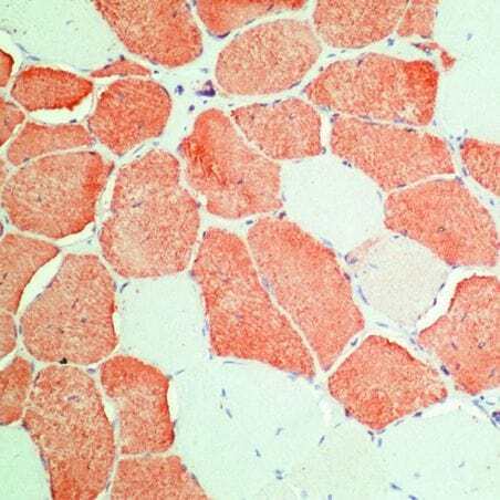
- Formalin-fixed, paraffin-embedded human skeletal muscle stained with Myosin, skeletal muscle antibody using peroxidase-conjugate and AEC chromogen. Note cytoplasmic staining of muscle cells.
Supportive validation
- Submitted by
- Invitrogen Antibodies (provider)
- Main image

- Experimental details
- Figure 2 Efficient fusion requires myomaker expression in both fusing cells but myomerger in one fusing cell. ( a ) Diagram showing the cell mixing approach to assess fusion between the populations of fibroblasts. Co-localization of GFP and NLS-TdTomato (NLS-Tom) in the nucleus represents fusion (arrows). Representative images demonstrate fusion of myomaker + myomerger + GFP + fibroblasts with myomaker + NLS-Tom + fibroblasts but not myomerger + NLS-Tom + fibroblasts. ( b ) Quantification of the percent of GFP + NLS-Tom + syncytia and the percent of nuclei in syncytia ( n =3). Dotted line on right panel represents fusion achieved when both cells express both myomaker and myomerger (from Fig. 1b ). ( c ) Heterologous fusion experiment between C2C12 myoblasts and GFP + fibroblasts infected with either empty, myomaker or myomerger. Representative immunofluorescent images to visualize co-localization of myosin and GFP (arrows), indicating fusion. Quantification of the percentage of GFP + myosin + cells ( n =3). Data are presented as mean+-s.e.m. * P

- Submitted by
- Invitrogen Antibodies (provider)
- Main image

- Experimental details
- Figure 4 Requirement of myomerger for myoblast fusion in vitro . ( a ) Immunoblotting for myomerger in WT and myomerger KO C2C12 cells on day 2 of differentiation. GAPDH was used as a loading control. ( b ) Representative immunofluorescence images on day 2 and day 4 of differentiation for WT and myomerger KO C2C12 cells. Myomerger KO cells differentiate but fail to fuse. ( c ) Quantification of the differentiation index, the percentage of nuclei in myosin + cells ( n =4). NS, not significant. ( d ) The percentage of myosin + cells that contain 1-2, 3-8, or >=9 nuclei after 4 days of differentiation, as an indicator of fusogenicity ( n =3). ( e ) qRT-PCR for the indicated myogenic transcripts ( n =4). Data are presented as mean+-s.e.m. * P

- Submitted by
- Invitrogen Antibodies (provider)
- Main image
- Experimental details
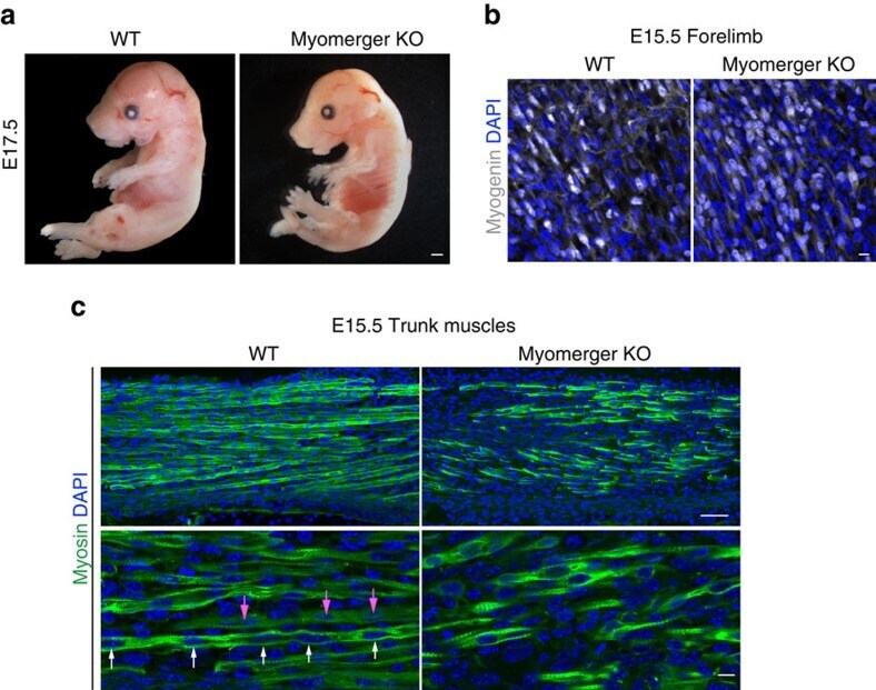
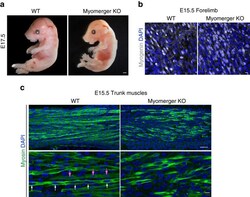
- Figure 6 Myomerger is essential for myoblast fusion and muscle formation during embryonic development. ( a ) Representative whole-mount images of WT and myomerger KO E17.5 embryos showing improper skeletal muscle formation in KO embryos ( n =4). ( b ) Immunofluorescence images for myogenin from WT and myomerger KO E15.5 forelimbs demonstrating that myomerger is not required for myogenic activation ( n =3). ( c ) Myosin immunofluorescence on the indicated E15.5 trunk muscles ( n =3). Multi-nucleated myofibres (arrows of same colour show nuclei within one myofibre) were observed in WT sections. Myomerger KO myocytes were myosin + with sarcomeres but remained mono-nucleated. Scale bars, 1 mm ( a ), 50 mum ( c ), top panels, 10 mum ( b , c ), bottom panels.