Explore
 Validate
 Learn
Learn Western blot
Antibody data
- Antibody Data
- Antigen structure
- References [0]
- Comments [0]
- Validations
- Western blot [3]
- Immunocytochemistry [3]
Submit
Validation data
Reference
Comment
Report error
- Product number
- PA1-10001 - Provider product page

- Provider
- Invitrogen Antibodies
- Product name
- NEFM Polyclonal Antibody
- Antibody type
- Polyclonal
- Antigen
- Purifed from natural sources
- Concentration
- Conc. Not Determined
No comments: Submit comment
Supportive validation
- Submitted by
- Invitrogen Antibodies (provider)
- Main image
- Experimental details
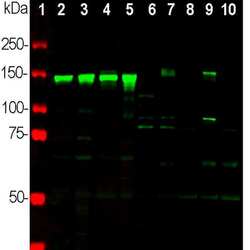
- Western blot analysis of NEFM in neuronal tissues and cell lysates using an NEFM polyclonal antibody (Product # PA1-10001) at a dilution 1:2,000 as seen in green. 1) protein standard (red), 2) rat brain 3) rat spinal cord, 4) mouse brain, 5) mouse spinal cord, 6) NIH/3T3 cells, 7) HEK293, 8) HeLa, 9) SH-SY5Y, and 10) C6 cells. Strong band at 145 kDa corresponds to rodent NEFM, and about 160 kDa band corresponds to human NEFM protein, visible in SHSY-5Y and HEK293 cells which have neuronal properties. NEFM is not expressed in HeLa and other cell lines tested.

- Submitted by
- Invitrogen Antibodies (provider)
- Main image
- Experimental details
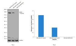
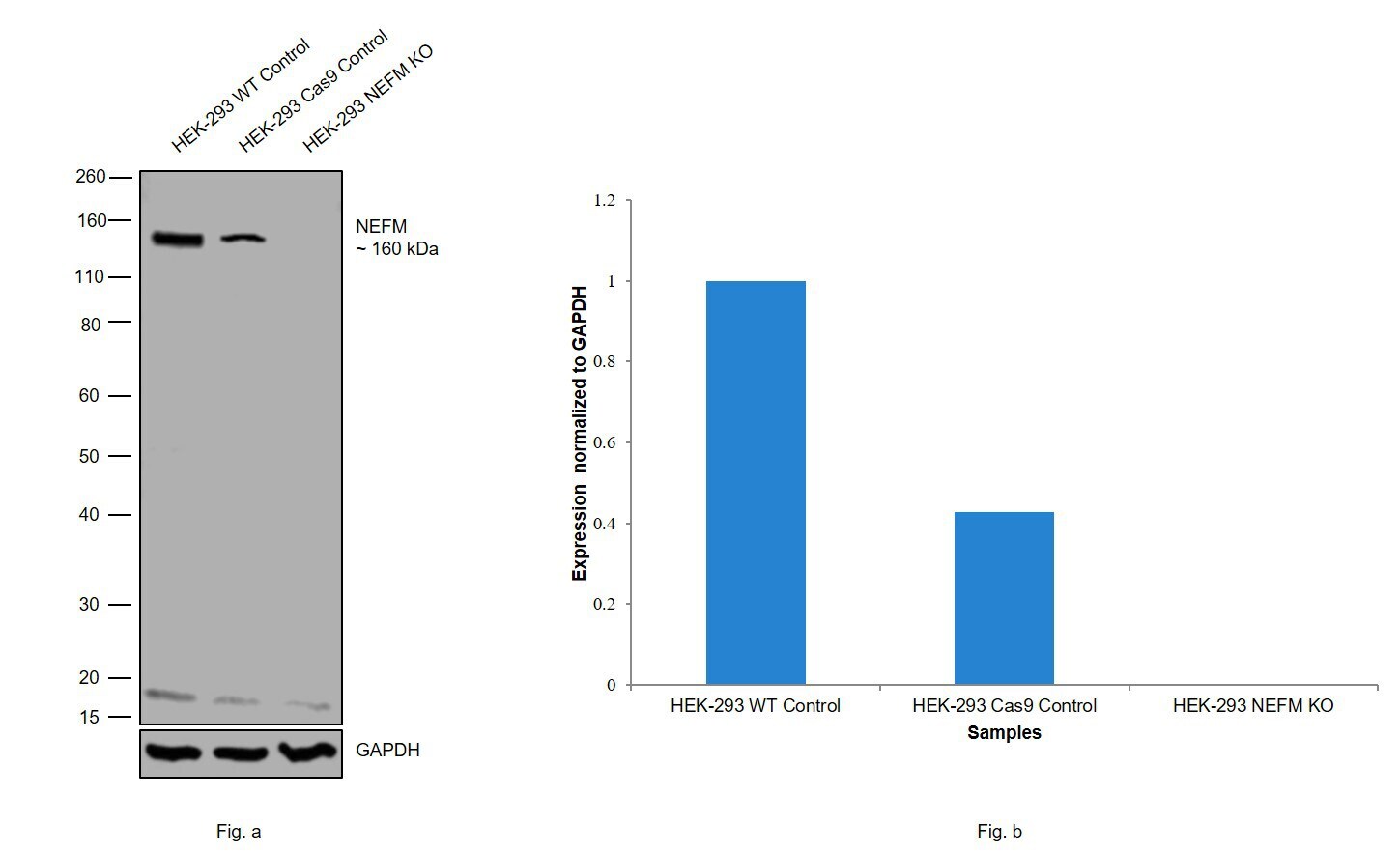
- Knockout of NEFM was achieved by CRISPR-Cas9 genome editing. Western blot analysis of NEFM was performed by loading 20 µg of HEK-293 wild type (Lane 1), HEK-293 Cas9 control (Lane 2), HEK-293 NEFM knockout (Lane 3) membrane enriched cell extracts. The blot was probed with Anti-NEFM Polyclonal Antibody (Product # PA1-10001) using 1:1000 dilution and Goat anti-Chicken IgY (H+L) Secondary Antibody, HRP (Product # A16054) using 1:4000 dilution). Loss of signal upon CRISPR mediated knockout (KO) confirms that antibody is specific to NEFM. Uncharacterised bands were observed at ~18 kDa.
- Submitted by
- Invitrogen Antibodies (provider)
- Main image
- Experimental details
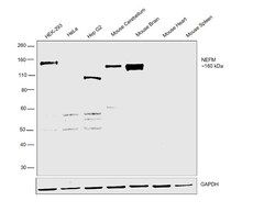
- Western blot was performed using Anti-NEFM Polyclonal Antibody (Product # PA1-10001) and a 160 kDa band corresponding to NEFM was observed in HEK-293 cell line, Mouse Cerebellum but not in HeLa, Hep G2 cell line, Mouse Heart and Mouse Spleen. Membrane enriched extracts (30 µg lysate) of HEK-293 (Lane 1), HeLa (Lane 2), Hep G2 (Lane 3), Mouse Cerebellum (Lane 4), Mouse Brain (Lane 5), Mouse Heart (Lane 6) and Mouse Spleen (Lane 7) were electrophoresed using Novex® NuPAGE® 4-12 % Bis-Tris gel (Product # NP0321BOX). Resolved proteins were then transferred onto a nitrocellulose membrane (Product # IB23001) by iBlot® 2 Dry Blotting System (Product # IB21001). The blot was probed with the primary antibody (1:2000 dilution) and detected by chemiluminescence with Goat anti-Chicken IgY (H+L) Secondary Antibody, HRP (Product # A16054, 1:4000 dilution) using the iBright FL 1000 (Product # A32752). Chemiluminescent detection was performed using Novex® ECL Chemiluminescent Substrate Reagent Kit (Product # WP20005).

Supportive validation
- Submitted by
- Invitrogen Antibodies (provider)
- Main image
- Experimental details
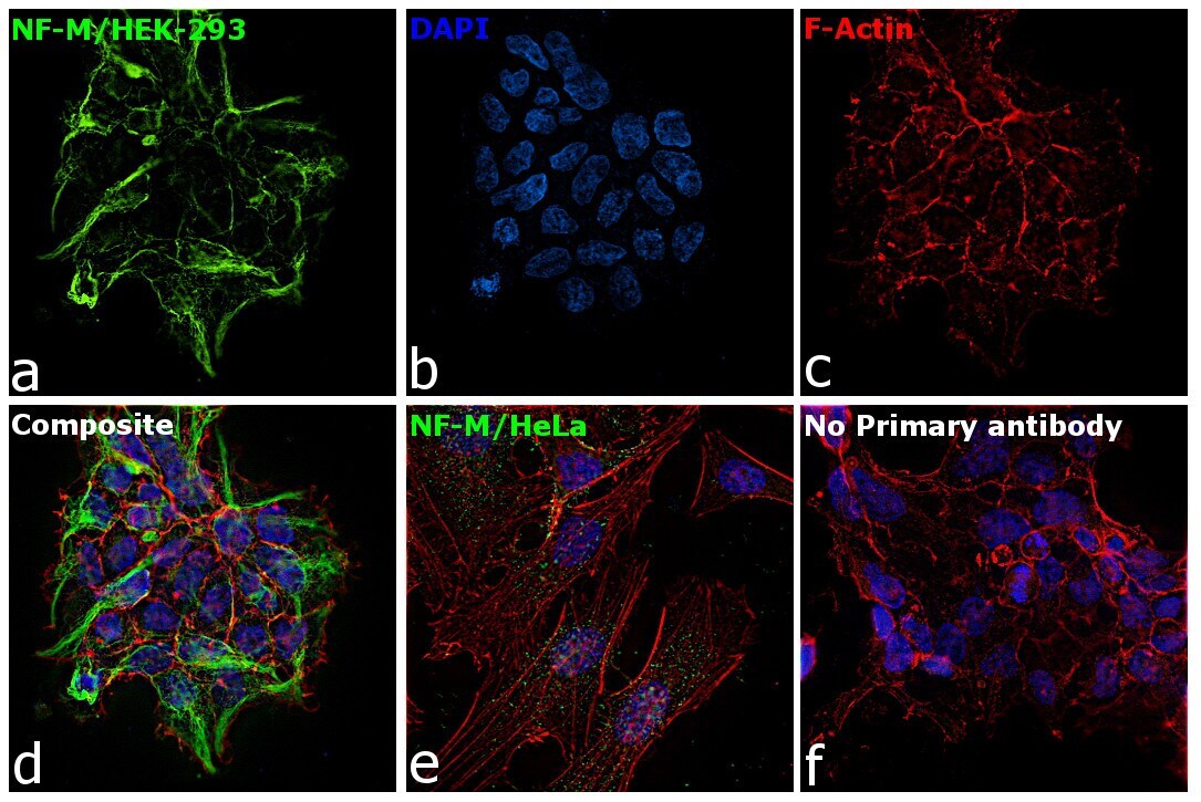
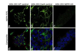
- Knockout of NEFM was achieved by CRISPR-Cas9 genome editing. Immunofluorescence analysis was performed on wild type HEK-293 cells (panel a,d), HEK-293 Cas9 cells (panels b,e) and HEK-293 NEFM KO cells (panel c,f). Cells were fixed, permeabilized, and labelled with NEFM Polyclonal Antibody (Product # PA1-10001) at 1:1000 dilution, followed by Goat anti-Chicken IgY (H+L) Secondary Antibody, Alexa Fluor® 488 conjugate (Product # A-11039) at a dilution of 1:2000. Nuclei (blue) were stained using ProLong™ Diamond Antifade Mountant with DAPI (Product # P36962), and Rhodamine Phalloidin (Product # R415) at a dilution of 1:300 was used for cytoskeletal F-actin (red) staining. Loss of signal (panel c,f) upon CRISPR mediated knockout (KO) confirms that antibody is specific to NEFM (green). The images were captured at 60X magnification.

- Submitted by
- Invitrogen Antibodies (provider)
- Main image

- Experimental details
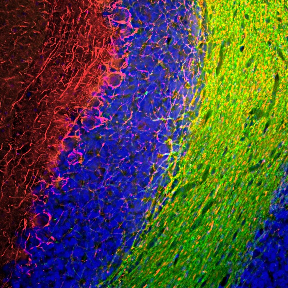
- Immunofluorescent analysis of NEFM in rat cerebellum. The rat cerebellum section was obtained following transcardial perfusion of the rat with 4% paraformaldehyde, brain was post fixed for 24 hours, and cut to 45µM. Free-floating sections were stained with an NEFM polyclonal antibody (Product # PA1-10001) at a dilution of 1:1,000 as seen in red, and costained with a CNPase monoclonal antibody at a dilution of 1:500 as seen in green, and with DAPI staining the nuclear DNA in blue. The NF-M antibody labels the axons of basket cells and other neurons, while the CNP antibody stains oligodendrocytes, cells that form the myelin sheathes around axons.
- Submitted by
- Invitrogen Antibodies (provider)
- Main image
- Experimental details
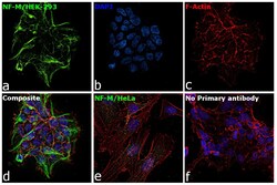
- Immunofluorescence analysis of NF-M was performed using 70% confluent log phase HEK-293 and HeLa cells. The cells were fixed with 4% paraformaldehyde for 10 minutes, permeabilized with 0.1% Triton™ X-100 for 15 minutes, and blocked with 2% BSA for 1 hour at room temperature. HEK-293 cells were labeled with NF-M Chicken Polyclonal Antibody (Product # PA1-10001) at 1:1000 dilution in 0.1% BSA, incubated at 4 degree Celsius overnight and then labeled with Goat anti-Chicken IgY (H+L) Secondary Antibody, Alexa Fluor® 488 conjugate (Product # A-11039) at a dilution of 1:2000 for 45 minutes at room temperature (Panel a: green). Nuclei (Panel b: blue) were stained with ProLong™ Diamond Antifade Mountant with DAPI (Product # P36962). F-actin (Panel c: red) was stained with Rhodamine Phalloidin (Product # R415). Panel d represents the merged image of HEK-293 showing cytoskeletal (intermediate filament) localization. Panel e represents the merged image of HeLa cells showing no expression for NF-M protein. Panel f represents control cells with no primary antibody to assess background. The images were captured at 60X magnification.