 Explore
 Validate
 Learn
Learn Immunocytochemistry
 Flow cytometry
Antibody data
- Antibody Data
- Antigen structure
- References [3]
- Comments [0]
- Validations
- Immunocytochemistry [1]
Submit
Validation data
Reference
Comment
Report error
- Product number
- ALX-804-010F-T100 - Provider product page

- Provider
- Enzo Life Sciences
- Product name
- FasL (mouse) monoclonal antibody (H11) (FITC conjugate)
- Antibody type
- Monoclonal
- Antigen
- Synthetic peptide
- Reactivity
- Mouse
- Host
- Rat
- Conjugate
- Green dye
- Isotype
- IgG
- Antibody clone number
- H11
- Vial size
- 100 tests
- Storage
- +4°C
- Handling
- Avoid freeze/thaw cycles.
Submitted references Transgenic expression of CD95 ligand on thyroid follicular cells confers immune privilege upon thyroid allografts.
CD4+ T cells reactivated with superantigen are both more sensitive to FasL-mediated killing and express a higher level of FasL.
Characterization of the non-functional Fas ligand of gld mice.
Tourneur L, Malassagne B, Batteux F, Fabre M, Mistou S, Lallemand E, Lores P, Chiocchia G
Journal of immunology (Baltimore, Md. : 1950) 2001 Aug 1;167(3):1338-46
Journal of immunology (Baltimore, Md. : 1950) 2001 Aug 1;167(3):1338-46
CD4+ T cells reactivated with superantigen are both more sensitive to FasL-mediated killing and express a higher level of FasL.
Wang JK, Zhu B, Ju ST, Tschopp J, Marshak-Rothstein A
Cellular immunology 1997 Aug 1;179(2):153-64
Cellular immunology 1997 Aug 1;179(2):153-64
Characterization of the non-functional Fas ligand of gld mice.
Hahne M, Peitsch MC, Irmler M, Schröter M, Lowin B, Rousseau M, Bron C, Renno T, French L, Tschopp J
International immunology 1995 Sep;7(9):1381-6
International immunology 1995 Sep;7(9):1381-6
No comments: Submit comment
Supportive validation
- Submitted by
- Enzo Life Sciences (provider)
- Main image
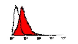
- Experimental details
- Flow cytometric profile of 293T cells transiently transfected with a FasL-expression plasmid (filled profile). The open profile corresponds to mock-transfected 293T cells. Method: FasL-transfected 293T cells (5 x 10E5) were incubated on ice for 30 min in 50 µl FACS buffer (PBS, 5% Fetal calf serum, 0.02% azide) containing 1µg of FITC-labelled H11 antibody. After washing in FACS buffer, cells were anlyzed by flow cytometry. Do not exclude pre-apoptotic cells during data acquisition (see T. Renno, M. Hahne, J. Tschopp and H. R. MacDonald, J. Exp. Med., 183, 431 (1996)). Note: Activated primary T cells do not express high levels of surface FasL!
