Explore
 Validate
 Learn
LearnMAB172-100
antibody from Novus Biologicals
Targeting: CXCR4
CD184, D2S201E, fusin, HM89, HSY3RR, LESTR, NPY3R, NPYR, NPYY3R
 Immunohistochemistry
Antibody data
- Antibody Data
- Antigen structure
- References [0]
- Comments [0]
- Validations
- Immunohistochemistry [2]
- Flow cytometry [1]
- Blocking/Neutralizing [1]
Submit
Validation data
Reference
Comment
Report error
- Product number
- MAB172-100 - Provider product page

- Provider
- Novus Biologicals
- Product name
- Mouse Monoclonal CXCR4 Antibody
- Antibody type
- Monoclonal
- Description
- Protein A or G purified from hybridoma culture supernatant. Detects human CXCR4. It will also react with cells expressing feline CXCR4 but not rat CXCR4.
- Reactivity
- Human
- Host
- Mouse
- Conjugate
- Unconjugated
- Isotype
- IgG
- Vial size
- 100 ug
- Concentration
- LYOPH
- Storage
- Use a manual defrost freezer and avoid repeated freeze-thaw cycles. 12 months from date of receipt, -20 to -70 degreesC as supplied. 1 month, 2 to 8 degreesC under sterile conditions after reconstitution. 6 months, -20 to -70 degreesC under sterile conditions after reconstitution.
No comments: Submit comment
Supportive validation
- Submitted by
- Novus Biologicals (provider)
- Main image
- Experimental details
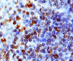
- CXCR4 in Human Spleen. CXCR4 was detected in immersion fixed paraffin-embedded sections of human spleen using Mouse Anti-Human CXCR4 Monoclonal Antibody (Catalog # MAB172) at 15 µg/mL overnight at 4 °C. Before incubation with the primary antibody, tissue was subjected to heat-induced epitope retrieval using Antigen Retrieval Reagent-Basic (Catalog # CTS013). Tissue was stained using the Anti-Mouse HRP-DAB Cell & Tissue Staining Kit (brown; Catalog # CTS002) and counterstained with hematoxylin (blue). Specific staining was localized to cytoplasm and nuclei. View our protocol for Chromogenic IHC Staining of Paraffin-embedded Tissue Sections.
- Submitted by
- Novus Biologicals (provider)
- Main image

- Experimental details
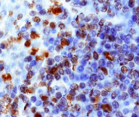
- CXCR4 in Human Lymph Node. CXCR4 was detected in immersion fixed paraffin-embedded sections of human lymph node using 15 µg/mL Human CXCR4 Monoclonal Antibody (Catalog # MAB172) overnight at 4 °C. Tissue was stained with the Anti-Mouse HRP-AEC Cell & Tissue Staining Kit (red; Catalog # CTS003) and counterstained with hematoxylin (blue). View our protocol for Chromogenic IHC Staining of Paraffin-embedded Tissue Sections.

Supportive validation
- Submitted by
- Novus Biologicals (provider)
- Main image
- Experimental details
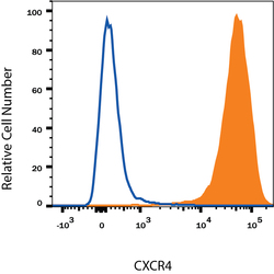
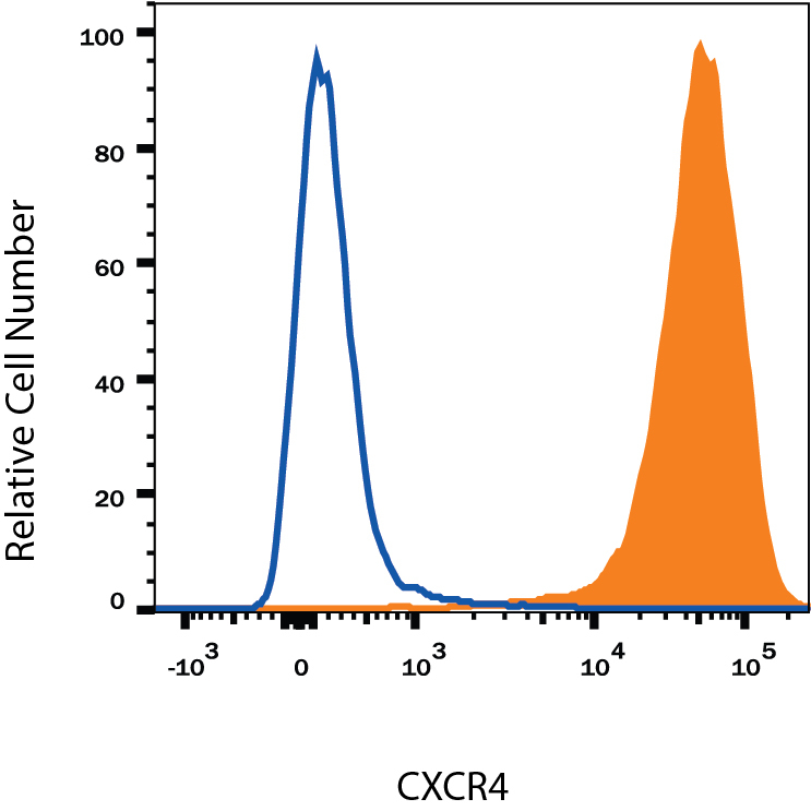
- Detection of CXCR4 in Jurkat Human Cell Line by Flow Cytometry. Jurkat human acute T cell leukemia cell line was stained with Mouse Anti-Human CXCR4 Monoclonal Antibody (Catalog # MAB172, filled histogram) or isotype control antibody (Catalog # MAB004, open histogram), followed by Allophycocyanin-conjugated Anti-Mouse IgG Secondary Antibody (Catalog # F0101B). View our protocol for Staining Membrane-associated Proteins.
Supportive validation
- Submitted by
- Novus Biologicals (provider)
- Main image

- Experimental details
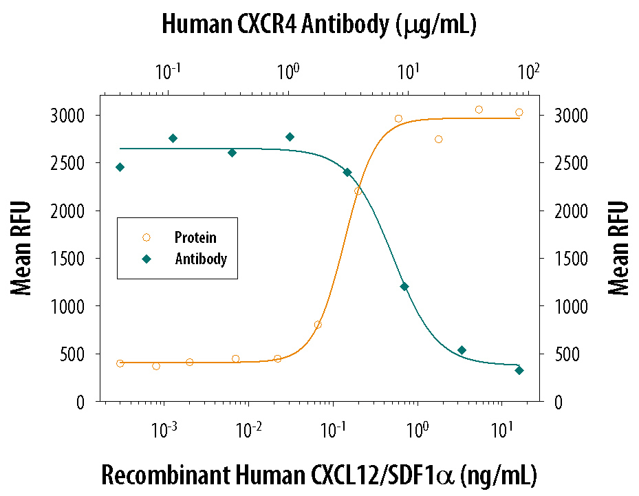
- Chemotaxis Induced by CXCL12/SDF-1 alpha and Neutralization by Human CXCR4 Antibody. Recombinant Human/Feline/Rhesus Macaque CXCL12/SDF-1 alpha (Catalog # 350-NS) chemoattracts the BaF3 mouse pro-B cell line transfected with human CXCR4 in a dose-dependent manner (orange line). The amount of cells that migrated through to the lower chemotaxis chamber was measured by Resazurin (Catalog # AR002). Chemotaxis elicited by Recombinant Human/Feline/Rhesus Macaque CXCL12/SDF-1 alpha (1 ng/mL) is neutralized (green line) by increasing concentrations of Mouse Anti-Human CXCR4 Monoclonal Antibody (Catalog # MAB172). The ND50 is typically 2.5-12 µg/mL.