Explore
 Validate
 Learn
Learn13-7096-81
antibody from Invitrogen Antibodies
Targeting: CCL2
GDCF-2, HC11, MCAF, MCP-1, MCP1, MGC9434, SCYA2, SMC-CF
 ELISA
ELISA Flow cytometry
Antibody data
- Antibody Data
- Antigen structure
- References [9]
- Comments [0]
- Validations
- Flow cytometry [1]
Submit
Validation data
Reference
Comment
Report error
- Product number
- 13-7096-81 - Provider product page

- Provider
- Invitrogen Antibodies
- Product name
- Anti-CCL2 (MCP-1) Monoclonal Antibody (2H5), Biotin, eBioscience™
- Antibody type
- Monoclonal
- Antigen
- Other
- Description
- Description: The 2H5 monoclonal antibody reacts with mouse, rat, and human monocyte chemoattractant protein-1 (MCP-1), also known as CCL2 and MCAF. Applications Reported: The 2H5 antibody has been reported for use in ELISA, intracellular staining for flow cytometric analysis, and cytokine neutralization. (Please use Functional Grade purified 2H5, cat. 16-7096, in functional assays.). Applications Tested: Mouse MCP-1 ELISA: The biotinylated 2H5 antibody has been tested as the detection antibody in a sandwich ELISA for analysis of mouse MCP-1 in combination with the affinity purified 4E2/MCP (14-7091) antibody for capture and recombinant mouse MCP-1 as the standard. A suitable range of concentrations of this antibody for ELISA detection is 0.5-2.0 µg/mL. A standard curve consisting of doubling dilutions of the recombinant standard over the range of 2000 pg/mL - 15 pg/mL should be included in each ELISA plate. Human MCP-1 ELISA: The biotinylated 2H5 antibody has been tested as the detection antibody in a sandwich ELISA for analysis of human MCP-1 in combination with the affinity purified 5D3-F7 (14-7099) antibody for capture and recombinant human MCP-1 as the standard. A suitable range of concentrations of this antibody for ELISA detection is 0.5-2.0 µg/mL. A standard curve consisting of doubling dilutions of the recombinant standard over the range of 2000 pg/mL - 15 pg/mL should be included in each ELISA plate. The Functional Grade purified 2H5 antibody has been tested for neutralization of MCP-1 bioactivity.The fluorochrome-conjugated 2H5 antibody has been tested for intracellular staining and flow cytometric analysis of mouse, rat, and human cells and can be used at less than or equal to 0.5 µg per test. A test is defined as the amount (µg) of antibody that will stain a cell sample in a final volume of 100 µL. Cell number should be determined empirically but can range from 10^5 to 10^8 cells/test. It is recommended that the antibody be carefully titrated for optimal performance in the assay of interest. Filtration: 0.2 µm post-manufacturing filtered.
- Reactivity
- Human, Mouse, Rat
- Conjugate
- Biotin
- Isotype
- IgG
- Antibody clone number
- 2H5
- Vial size
- 50 µg
- Concentration
- 0.5 mg/mL
- Storage
- 4° C, store in dark, DO NOT FREEZE!
Submitted references Development and validation of an immunosensor for monocyte chemotactic protein 1 using a silicon photonic microring resonator biosensing platform.
Conditioned medium from umbilical cord mesenchymal stem cells induces migration and angiogenesis.
Oxidized low-density lipoprotein induces long-term proinflammatory cytokine production and foam cell formation via epigenetic reprogramming of monocytes.
The liver X receptor agonist GW3965 improves recovery from mild repetitive traumatic brain injury in mice partly through apolipoprotein E.
CCR2 defines a distinct population of NK cells and mediates their migration during influenza virus infection in mice.
Synergistic drug combinations tend to improve therapeutically relevant selectivity.
Mechanisms underlying renoprotection during renin-angiotensin system blockade.
Mechanisms underlying renoprotection during renin-angiotensin system blockade.
Macrophage inflammatory protein-2 and KC induce chemokine production by mouse astrocytes.
Valera E, Shia WW, Bailey RC
Clinical biochemistry 2016 Jan;49(1-2):121-6
Clinical biochemistry 2016 Jan;49(1-2):121-6
Conditioned medium from umbilical cord mesenchymal stem cells induces migration and angiogenesis.
Shen C, Lie P, Miao T, Yu M, Lu Q, Feng T, Li J, Zu T, Liu X, Li H
Molecular medicine reports 2015 Jul;12(1):20-30
Molecular medicine reports 2015 Jul;12(1):20-30
Oxidized low-density lipoprotein induces long-term proinflammatory cytokine production and foam cell formation via epigenetic reprogramming of monocytes.
Bekkering S, Quintin J, Joosten LA, van der Meer JW, Netea MG, Riksen NP
Arteriosclerosis, thrombosis, and vascular biology 2014 Aug;34(8):1731-8
Arteriosclerosis, thrombosis, and vascular biology 2014 Aug;34(8):1731-8
The liver X receptor agonist GW3965 improves recovery from mild repetitive traumatic brain injury in mice partly through apolipoprotein E.
Namjoshi DR, Martin G, Donkin J, Wilkinson A, Stukas S, Fan J, Carr M, Tabarestani S, Wuerth K, Hancock RE, Wellington CL
PloS one 2013;8(1):e53529
PloS one 2013;8(1):e53529
CCR2 defines a distinct population of NK cells and mediates their migration during influenza virus infection in mice.
van Helden MJ, Zaiss DM, Sijts AJ
PloS one 2012;7(12):e52027
PloS one 2012;7(12):e52027
Synergistic drug combinations tend to improve therapeutically relevant selectivity.
Lehár J, Krueger AS, Avery W, Heilbut AM, Johansen LM, Price ER, Rickles RJ, Short GF 3rd, Staunton JE, Jin X, Lee MS, Zimmermann GR, Borisy AA
Nature biotechnology 2009 Jul;27(7):659-66
Nature biotechnology 2009 Jul;27(7):659-66
Mechanisms underlying renoprotection during renin-angiotensin system blockade.
Taal MW, Chertow GM, Rennke HG, Gurnani A, Jiang T, Shahsafaei A, Troy JL, Brenner BM, Mackenzie HS
American journal of physiology. Renal physiology 2001 Feb;280(2):F343-55
American journal of physiology. Renal physiology 2001 Feb;280(2):F343-55
Macrophage inflammatory protein-2 and KC induce chemokine production by mouse astrocytes.
Luo Y, Fischer FR, Hancock WW, Dorf ME
Journal of immunology (Baltimore, Md. : 1950) 2000 Oct 1;165(7):4015-23
Journal of immunology (Baltimore, Md. : 1950) 2000 Oct 1;165(7):4015-23
No comments: Submit comment
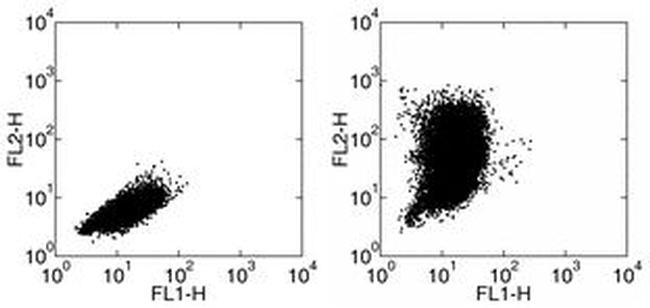
Supportive validation
- Submitted by
- Invitrogen Antibodies (provider)
- Main image
- Experimental details
- Staining of mouse thiogylcolate induced peritoneal exudate cells (PECs) stimulated with LPS in the presence of Brefeldin A for 24 hours. with Anti-mouse F4/80 FITC (Product # 11-4801-82) followed by intracellular staining with Rat IgG2a Isotype Control PE (Product # 12-4321-80) (left) or Anti-Mouse CCL2 PE.
- Conjugate
- Biotin