 Explore
 Validate
 Learn
Learn Western blot
Antibody data
- Antibody Data
- Antigen structure
- References [0]
- Comments [0]
- Validations
- Western blot [1]
- Immunocytochemistry [1]
Submit
Validation data
Reference
Comment
Report error
- Product number
- M807 - Provider product page

- Provider
- Invitrogen Antibodies
- Product name
- EGF Monoclonal Antibody (9A1)
- Antibody type
- Monoclonal
- Antigen
- Recombinant full-length protein
- Description
- The M807 anti-EGF antibody has successfully been paired as the detection antibody in a sandwich ELISA with coating antibody MA805 (Clone 1H11).
- Antibody clone number
- 9A1
- Concentration
- 1 mg/mL
No comments: Submit comment
Supportive validation
- Submitted by
- Invitrogen Antibodies (provider)
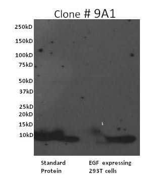
- Main image

- Experimental details
- Western blot analysis of Human EGF was performed by loading 2 µg of recombinant human EGF (Lane 1) or supernatant from an EGF expression clone transfected in 293T cells onto a 4-20% Tris-HCl polyacrylamide gel. Proteins were transferred to a PVDF membrane and blocked with 5% Milk/TBST for at least 1 hour. Membranes were probed with an EGF monoclonal antibody recognizing Human EGF (Product # M807) at a dilution of 1:5000 overnight at 4°C on a rocking platform. Membranes were washed in TBS-0.1%Tween 20 and probed with a goat anti-rabbit-HRP secondary antibody (Product # 31430) at a dilution of 1:10,000 for at least one hour. Membranes were washed and chemiluminescent detection performed using Super Signal West Dura (Product # 34075).
Supportive validation
- Submitted by
- Invitrogen Antibodies (provider)
- Main image
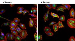
- Experimental details
- Immunofluorescent analysis of EGF (green) in HeLa cells either left serum starved (left panel) or treated with serum (right panel) for 30 minutes. Formalin fixed cells were permeabilized with 0.1% Triton X-100 in TBS for 10 minutes at room temperature and blocked with 1% Blocker BSA (Product # 37525) for 15 minutes at room temperature. Cells were probed with an EGF monoclonal antibody (Product # M807) at a dilution of 1:50 for at least 1 hour at room temperature, washed with PBS, and incubated with DyLight 488 goat anti-mouse IgG secondary antibody (Product # 35502) at a dilution of 1:400 for 30 minutes at room temperature. F-Actin (red) was stained with DyLight 554 Phalloidin (Product # 21834) and nuclei (blue) were stained with Hoechst 33342 dye (Product # 62249). Images were taken on a Thermo Scientific ArrayScan at 20X magnification.
