Explore
 Validate
 Learn
Learn Western blot
Antibody data
- Antibody Data
- Antigen structure
- References [0]
- Comments [0]
- Validations
- Western blot [1]
- Immunocytochemistry [1]
- Immunohistochemistry [1]
Submit
Validation data
Reference
Comment
Report error
- Product number
- AP11498PU-N - Provider product page

- Provider
- Acris Antibodies GmbH
- Proper citation
- Acris Antibodies GmbH Cat#AP11498PU-N, RRID:AB_1769959
- Product name
- anti NANOG (101-131)
- Antibody type
- Polyclonal
- Antigen
- This antibody is generated from rabbits immunized with a KLH conjugated synthetic peptide selected from the center region (aa101-131) of human NANOG.
- Reactivity
- Human
- Host
- Rabbit
- Vial size
- 0.4 ml
- Concentration
- lot specific
No comments: Submit comment
Supportive validation
- Submitted by
- Acris Antibodies GmbH (provider)
- Main image

- Experimental details
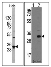
- (LEFT) Western blot analysis of NANOG Antibody (Center) (AP11498PU-N) in Hela cell line lysates (35 µg/lane). NANOG (arrow) was detected using the purified Pab (1:60 dilution). (RIGHT) Western blot analysis of NANOG (arrow) using rabbit polyclonal NANOG Antibody (Center) (AP11498PU-N). 293 cell lysates (2 µg/lane) either nontransfected (Lane 1) or transiently transfected with the NANOG gene (Lane 2) (Origene Technologies).
Supportive validation
- Submitted by
- Acris Antibodies GmbH (provider)
- Main image

- Experimental details
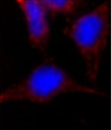
- Immunofluorescence analysis of NANOG Antibody (Center) (AP11498PU-N) in HeLa cells. 0.025 mg/ml primary antibody was followed by Alexa-Fluor-546-conjugated donkey anti-rabbit lgG (H+L). Alexa-Fluor-546 emits orange fluorescence. Blue counterstaining is DAPI.
Supportive validation
- Submitted by
- Acris Antibodies GmbH (provider)
- Main image

- Experimental details
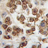
- Formalin-fixed and paraffin-embedded human testis tissue reacted with NANOG antibody (Center), which was peroxidase-conjugated to the secondary antibody, followed by DAB staining. This data demonstrates the use of this antibody for immunohistochemistry; clinical relevance has not been evaluated.